. 2022 Jun;19(3):617-628.
doi: 10.1007/s13770-021-00414-4. Epub 2021 Dec 28.

M2 Macrophage-Derived Concentrated Conditioned Media Significantly Improves Skin Wound Healing

Affiliations

M2 Macrophage-Derived Concentrated Conditioned Media Significantly Improves Skin Wound Healing

Cininta Savitri et al. Tissue Eng Regen Med. 2022 Jun.

Abstract

Background: Macrophages, with many different phenotypes play a major role during wound healing process, secreting the cytokines crucial to angiogenesis, cell recruitment and ECM remodeling. Therefore, macrophage-derived cytokines may be attractive therapeutic resource for wound healing.

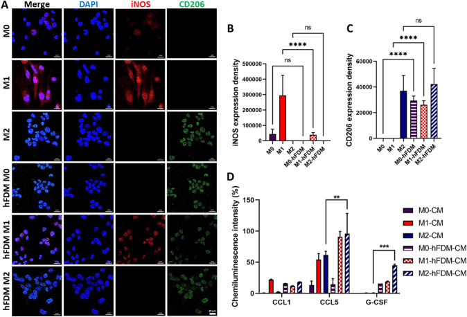
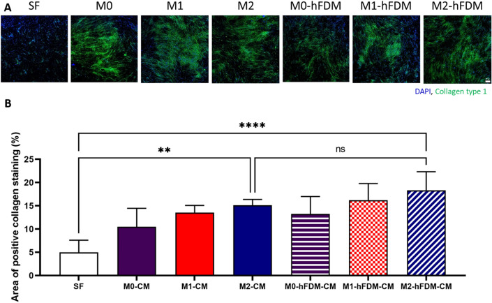
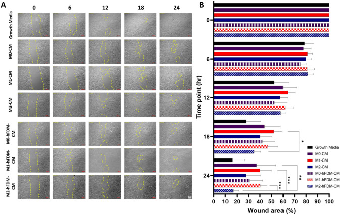
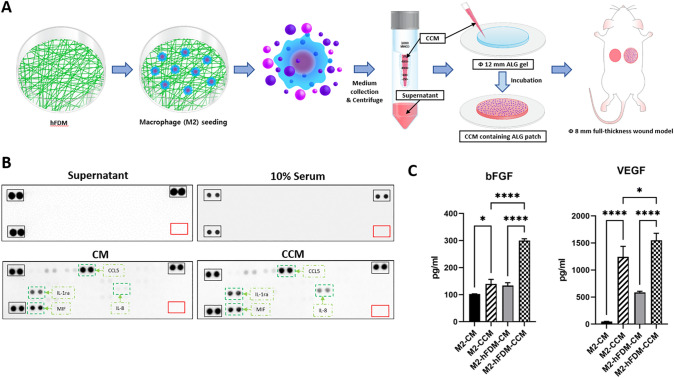
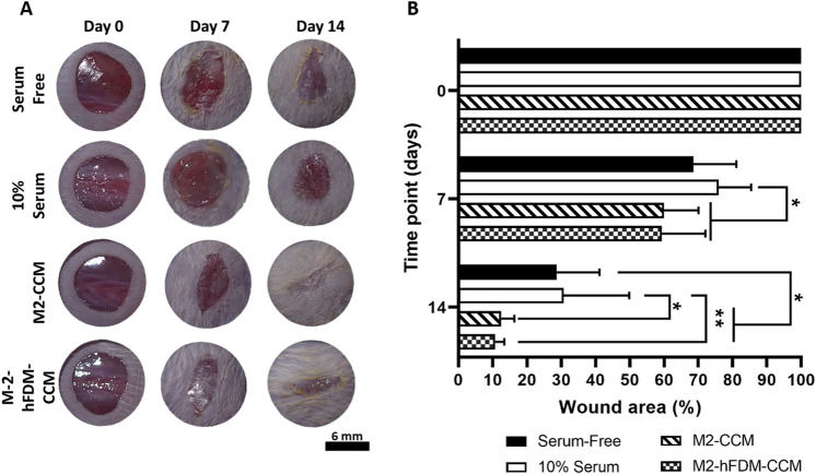
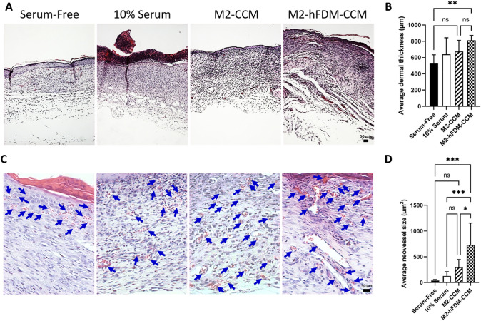
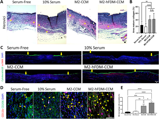
Methods: To obtain a conditioned media (CM) from macrophages, human monocyte THP-1 cells were seeded on TCP or human fibroblast-derived matrix (hFDM) and they were differentiated into M1 or M2 phenotype using distinct protocols. A combination of different substrates and macrophage phenotypes produced M1- and M2-CM or M1-hFDM- and M2-hFDM-CM, respectively. Proteome microarray determines the cytokine contents in those CMs. CMs-treated human dermal fibroblast (hDFB) was analyzed using collagen synthesis and wound scratch assay. Concentrated form of the CM (CCM), obtained by high-speed centrifugation, was administered to a murine full-thickness wound model using alginate patch, where alginate patch was incubated in the M2-CCM overnight at 4 °C before transplantation. On 14 day post-treatment, examination was carried out through H&E and Herovici staining. Keratinocyte and M2 macrophages were also evaluated via immunofluorescence staining.

Results: Cytokine analysis of CMs found CCL1, CCL5, and G-CSF, where CCL5 is more dominant. We found increased collagen synthesis and faster wound closure in hDFB treated with M2-CM. Full-thickness wounds treated by M2-hFDM-CCM containing alginate patch showed early wound closure, larger blood vessels, increased mature collagen deposition, enhanced keratinocyte maturation and more M2-macrophage population.

Conclusion: Our study demonstrated therapeutic potential of the CM derived from M2 macrophages, where the cytokines in the CM may have played an active role for enhanced wound healing.

Keywords: Alginate; Cytokines; Extracellular matrix; Macrophage; Wound healing.


Conflict of interest statement

There are no conflicts to declare.

Figures

Fig. 1
Fig. 1
Macrophage phenotype on different substrates (TCP and hFDM) and cytokine release of macrophage in vitro. Immunofluorescence staining of macrophage phenotypes (M1 and M2) surface marker: (A) CD206 (M2 marker) and iNOS (M1 marker). B, C Quantification of fluorescence signals of iNOS and CD206. D Proteomic microarray of macrophage-derived, six different conditioned media (CMs) presents wound healing-related cytokines, CCL1, CCL5, and G-CSF. Scale bar is 20 µm. Statistically significant difference (**p < 0.01, ***p < 0.001)
Fig. 2
Fig. 2
A Macrophage conditioned media can stimulate collagen deposition of hDFB in vitro. Immunofluorescence staining of collagen type I produced by hDFB. It shows M2-CM and M2-hFDM-CM could induce more collagen production of hDFB compared to that of serum-free medium. B Quantitative analysis of collagen production level of each CM-treated hDFB. SF represents serum-free media. Scale bar is 200 μm. Statistically significant difference (**p < 0.01, ****p < 0.0001)
Fig. 3
Fig. 3
A M2-hFDM-CM accelerates artificial wound closure in hDFB wound scratch test. Representative images of hDFB wound closure with time treated with different macrophage-derived CMs. The yellow marks indicate the wound still uncovered with hDFB. B Quantitative analysis of wound closure at specific time points. Scale bar is 200 μm. Statistically significant difference (*p < 0.05, **p < 0.01, ***p < 0.001)
Fig. 4
Fig. 4
High speed centrifugation successfully concentrated conditioned media. A Schematic of macrophage-derived concentrated CM (CCM) preparation and implantation of CCM-embedded alginate patch into skin wound model. B Proteomic microarray of cytokines contained in supernatant, 10% serum, CM and CCM, respectively. No signals detected in the supernatant. C ELISA analysis of angiogenic growth factors (bFGF and VEGF) contained in two different types of CM and CCM. (*p < 0.05, ****p < 0.0001)
Fig. 5
Fig. 5
CCM treated murine full-thickness skin wound shows faster wound closure. A Representative images of wound closure on day 0, 7, and 14 post-treatment of four test groups (serum-free, 10% serum, M2-CM and M2-hFDM-CM) (n = 6 each group), respectively. B Measurement of wound closure ratio (%) at specific time points. Scale bar is 6 mm. Statistically significant difference (*p < 0.05, **p < 0.01)
Fig. 6
Fig. 6
CCM-treated wounds reveal better histological outcome and advanced neovascularization. A Representative H&E staining images of the regenerated wound tissues on day 14 post-treatment. B Quantitative analysis of dermis thickness. C High magnification of H&E images shows the neovessels on day 14 post-treatment. Blue arrows mark neovessels. D Quantitative analysis of average vessel size. Scale bar is 50 μm. Statistically significant difference (*p < 0.05, **p < 0.01, ***p < 0.001)
Fig. 7
Fig. 7
Analysis of CCM-treated wounds via Herovici, keratinocytes and M2 macrophage staining, respectively. A Representative Herovici staining images of the regenerated wound tissues on day 14. Yellow asterisk denotes mature collagen in red tone. B Quantitative analysis of mature collagen area. C Representative cytokeratin10 and α-SMA staining images of the epidermis layer on day 14. Yellow arrow marks wound edges. D Representative CD11b and CD206 positive images on day 14. Yellow triangles denote double positive cells and white star symbols show blood vessels. E Quantification of positively double stained cells with CD11b and CD206 (M2 macrophages). Scale bar is 50 μm. Statistically significant difference (*p < 0.05, **p < 0.01, ***p < 0.001, ****p < 0.0001)

References

    1. Barrientos S, Stojadinovic O, Golinko MS, Brem H, Tomic-Canic M. Growth factors and cytokines in wound healing. Wound Repair Regener. 2008;16:585–601. doi: 10.1111/j.1524-475X.2008.00410.x. - DOI - PubMed
    1. Behm B, Babilas P, Landthaler M, Schreml S. Cytokines, chemokines and growth factors in wound healing. J Eur Acad Dermatol Venereol. 2012;26:812–820. doi: 10.1111/j.1468-3083.2011.04415.x. - DOI - PubMed
    1. Rodero MP, Khosrotehrani K. Skin wound healing modulation by macrophages. Int J Clin Exp Pathol. 2010;3:643–653. - PMC - PubMed
    1. van der Veer W, van Egmond M, Niessen F, Beelen R. Macrophages in skin injury and repair. Immunobiology. 2011;216:753–762. doi: 10.1016/j.imbio.2011.01.001. - DOI - PubMed
    1. Minutti CM, Knipper JA, Allen JE, Zaiss DM. Tissue-specific contribution of macrophages to wound healing. Semin Cell Dev Biol. 2017;61:3-11. - PubMed

Publication types

LinkOut - more resources