

Bilateral Sequential Paracentral Acute Middle Maculopathy
- PMID: 34963271
- PMCID: PMC8715649
- DOI: 10.4274/tjo.galenos.2021.50207
Bilateral Sequential Paracentral Acute Middle Maculopathy
Abstract
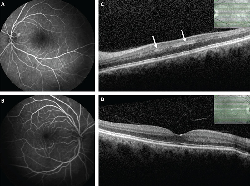
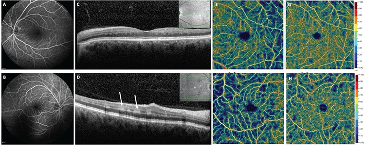
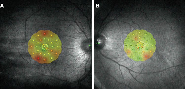
We aim to present a case with bilateral sequential paracentral acute middle maculopathy (PAMM). A 57-year-old man presented with paracentral scotoma in the left eye. The patient's multimodal imaging findings were consistent with PAMM in the left eye. Extensive systemic work-up revealed hypertension and a history of cerebrovascular event. One year after initial presentation, the patient had a subsequent decrease in visual acuity in the right eye and developed optical coherence tomography findings consistent with PAMM, whereas the left eye showed resolved PAMM findings. Although rare, PAMM can occur bilaterally. Clinicians should monitor unilateral PAMM patients with systemic vasculopathy for involvement in the fellow eye.
Keywords: PAMM; cerebrovascular event; hypertension; paracentral acute middle maculopathy.
Conflict of interest statement
Figures
References
-
- Sarraf D, Rahimy E, Fawzi AA, Sohn E, Barbazetto I, Zacks DN, Mittra RA, Klancnik JM Jr, Mrejen S, Goldberg NR, Beardsley R, Sorenson JA, Freund KB. Paracentral acute middle maculopathy: a new variant of acute macular neuroretinopathy associated with retinal capillary ischemia. JAMA Ophthalmol. 2013;131:1275–1287. - PubMed
-
- Iafe NA, Onclinx T, Tsui I, Sarraf D. Paracentral acute middle maculopathy and deep retinal capillary plexus infarction secondary to reperfused central retinal artery occlusion. Retina Cases Brief Rep. 2017;11(Suppl1):S90–S93. - PubMed
-
- Rahimy E, Kuehlewein L, Sadda SR, Sarraf D. Paracentral acute middle maculopathy: What we knew then and what we know now. Retina. 2015;35:1921–1930. - PubMed
-
- Maltsev DS, Kulikov AN, Burnasheva MA, Chhablani J. Prevalence of resolved paracentral acute middle maculopathy lesions in fellow eyes of patients with unilateral retinal vein occlusion. Acta Ophthalmol. 2020;98:e22–e28. - PubMed
Publication types
MeSH terms
LinkOut - more resources
Full Text Sources
Medical