. 2021 Dec 28;19(1):453.
doi: 10.1186/s12951-021-01097-8.

Novel brain-targeted nanomicelles for anti-glioma therapy mediated by the ApoE-enriched protein corona in vivo

Affiliations

Novel brain-targeted nanomicelles for anti-glioma therapy mediated by the ApoE-enriched protein corona in vivo

Zhe-Ao Zhang et al. J Nanobiotechnology. .

Abstract

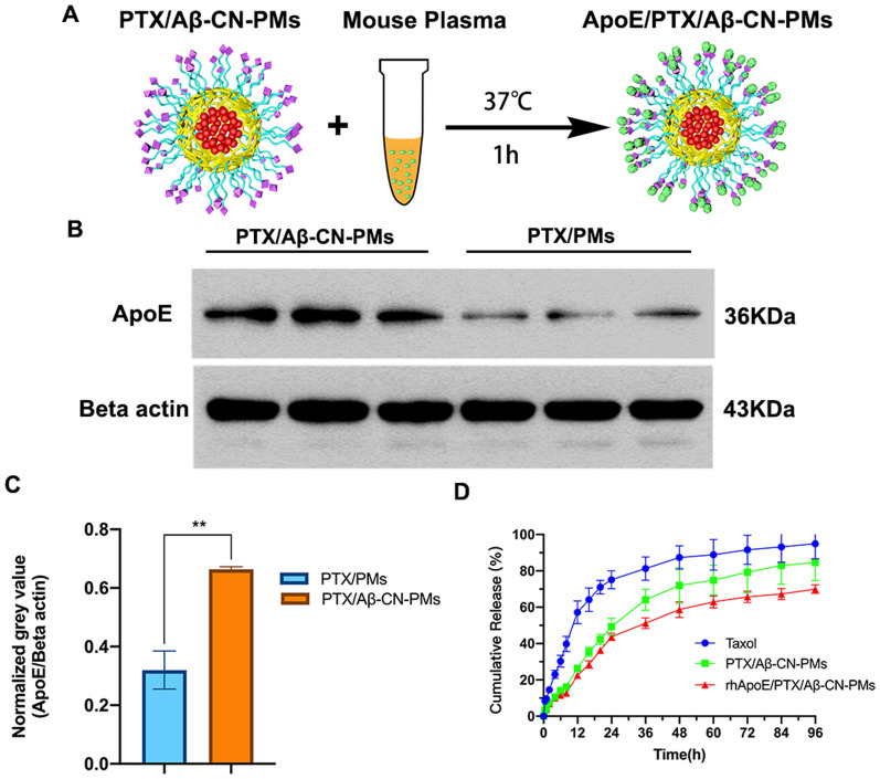
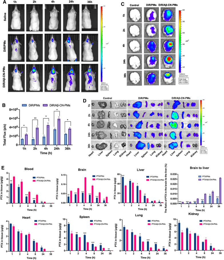
Background: The interactions between nanoparticles (NPs) and plasma proteins form a protein corona around NPs after entering the biological environment, which provides new biological properties to NPs and mediates their interactions with cells and biological barriers. Given the inevitable interactions, we regard nanoparticle‒protein interactions as a tool for designing protein corona-mediated drug delivery systems. Herein, we demonstrate the successful application of protein corona-mediated brain-targeted nanomicelles in the treatment of glioma, loading them with paclitaxel (PTX), and decorating them with amyloid β-protein (Aβ)-CN peptide (PTX/Aβ-CN-PMs). Aβ-CN peptide, like the Aβ1-42 peptide, specifically binds to the lipid-binding domain of apolipoprotein E (ApoE) in vivo to form the ApoE-enriched protein corona surrounding Aβ-CN-PMs (ApoE/PTX/Aβ-CN-PMs). The receptor-binding domain of the ApoE then combines with low-density lipoprotein receptor (LDLr) and LDLr-related protein 1 receptor (LRP1r) expressed in the blood-brain barrier and glioma, effectively mediating brain-targeted delivery.

Methods: PTX/Aβ-CN-PMs were prepared using a film hydration method with sonication, which was simple and feasible. The specific formation of the ApoE-enriched protein corona around nanoparticles was characterized by Western blotting analysis and LC-MS/MS. The in vitro physicochemical properties and in vivo anti-glioma effects of PTX/Aβ-CN-PMs were also well studied.

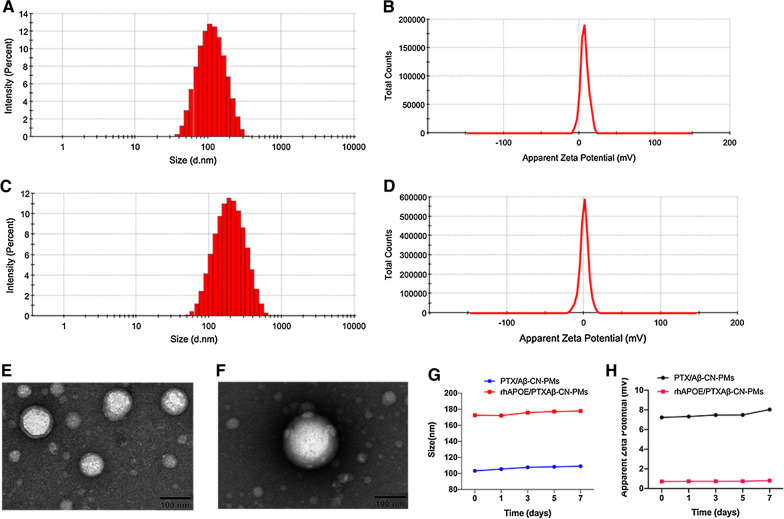
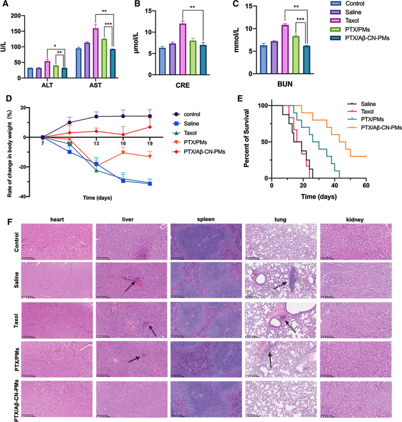
Results: The average size and zeta potential of PTX/Aβ-CN-PMs and ApoE/PTX/Aβ-CN-PMs were 103.1 nm, 172.3 nm, 7.23 mV, and 0.715 mV, respectively. PTX was efficiently loaded into PTX/Aβ-CN-PMs, and the PTX release from rhApoE/PTX/Aβ-CN-PMs exhibited a sustained-release pattern in vitro. The formation of the ApoE-enriched protein corona significantly improved the cellular uptake of Aβ-CN-PMs on C6 cells and human umbilical vein endothelial cells (HUVECs) and enhanced permeability to the blood-brain tumor barrier in vitro. Meanwhile, PTX/Aβ-CN-PMs with ApoE-enriched protein corona had a greater ability to inhibit cell proliferation and induce cell apoptosis than taxol. Importantly, PTX/Aβ-CN-PMs exhibited better anti-glioma effects and tissue distribution profile with rapid accumulation in glioma tissues in vivo and prolonged median survival of glioma-bearing mice compared to those associated with PMs without the ApoE protein corona.

Conclusions: The designed PTX/Aβ-CN-PMs exhibited significantly enhanced anti-glioma efficacy. Importantly, this study provided a strategy for the rational design of a protein corona-based brain-targeted drug delivery system. More crucially, we utilized the unfavorable side of the protein corona and converted it into an advantage to achieve brain-targeted drug delivery.

Keywords: ApoE protein corona; Glioma; Paclitaxel; Targeting therapy.


Conflict of interest statement

The authors declare no conflicts of interest in the paper.

Figures

Scheme 1
Scheme 1
A PTX loaded Aβ-CN-PMs (PTX/Aβ-CN-PMs) were prepared by film-hydration method with sonication. B PTX/Aβ-CN-PMs effectively captured ApoE and the brain targeted ApoE protein corona surrounding PTX/Aβ-CN-PMs crossed the BBB and BBTB, following by endocytosis into glioma cells.
Fig. 1
Fig. 1
Synthesis and characterization of mPEG2000-PLA1300, Mal-PEG2000-PLA1300 and Aβ-CN-PEG2000-PLA1300. A Synthesis route of mPEG2000-PLA1300 (A1), Mal- PEG2000-PLA1300 (A2), and Aβ-CN-PEG2000-PLA1300 (A3). B 1H-NMR spectra of mPEG2000-PLA1300 (B1), Mal-PEG2000-PLA1300 (B2), and Aβ-CN-PEG2000-PLA1300 (B3)
Fig. 2
Fig. 2
Characterizations of PTX/Aβ-CN-PMs and ApoE/PTX/Aβ-CN-PMs. A, C Size distribution of PTX/Aβ-CN-PMs and ApoE/PTX/Aβ-CN-PMs by dynamic light-scattering analysis. B, D Zeta potential distribution of PTX/Aβ-CN-PMs and ApoE/PTX/Aβ-CN-PMs. E, F TEM images of PTX/Aβ-CN-PMs and ApoE/PTX/Aβ-CN-PMs; scale bar = 100 nm. G The size stability of PTX/Aβ-CN-PMs and ApoE/PTX/Aβ-CN-PMs for seven days. H The zeta potential stability of PTX/Aβ-CN-PMs and ApoE/PTX/Aβ-CN-PMs for seven days
Fig. 3
Fig. 3
A Schematic diagram of PTX/Aβ-CN-PMs were incubated with the mouse plasma for 1 h at 37 °C in vitro. B Western Blotting results of the absorbed ApoE on PTX/PMs and PTX/Aβ-CN-PMs. C Gel Image system ver.4.00 (Tanon, China) was performed to quantitatively analyze the absorbed protein ApoE. means ± SD, n = 3; *P < 0.05, **P < 0.01, ***P < 0.001. D The PTX release profiles of taxol, PTX/Aβ-CN-PMs and rhApoE/PTX/Aβ-CN-PMs in sodium salicylate solution (1 M, pH 7.4)
Fig. 4
Fig. 4
Protein corona characterization. A The types and abundance of the adsorbed proteins on PTX/Aβ-CN-PMs and PTX/PMs. B The types and abundance of apolipoproteins on PTX/Aβ-CN-PMs and PTX/PMs
Fig. 5
Fig. 5
In vitro cytotoxicity study. In vitro cytotoxicity of blank Aβ-CN-PMs and rhApoE/Aβ-CN-PMs on C6 cells (A) and on HUVEC cells (B). C Inhibitory capacity of Taxol, PTX/Aβ-CN-PMs and rhApoE/PTX/Aβ-CN-PMs against C6 cells proliferation. Means ± SD, n = 3; *P < 0.05, **P < 0.01, ***P < 0.001 versus the rhApoE/PTX/Aβ-CN-PMs group
Fig. 6
Fig. 6
In vitro cellular uptake on C6 cells. A CLSM observation of C6 cells after treatment with free Cou-6, Cou-6/PMs, Cou-6/Aβ-CN-PMs, rhApoE/Cou-6/Aβ-CN-PMs for 15, 30, 60 and 120 min (magnification × 200), respectively. B, C Quantitative analysis of cellular uptake by flow cytometry after incubation with free Cou-6, Cou-6/PMs, Cou-6/Aβ-CN-PMs, rhApoE/Cou-6/Aβ-CN-PMs for 2 h, respectively. Means ± SD, n = 3; *P < 0.05, **P < 0.01, ***P < 0.001 versus rhApoE/Cou-6/Aβ-CN-PMs group. D, E Quantitative analysis of cellular uptake by flow cytometry after incubation with rhApoE/Cou-6/Aβ-CN-PMs for 15, 30, 60 and 120 min. F Cellular uptake analysis of rhApoE/Cou-6/Aβ-CN-PMs after incubation with different endocytic inhibitors by flow cytometry. Means ± SD, n = 3; *P < 0.05, **P < 0.01, ***P < 0.001 versus the control group without endocytic inhibitors
Fig. 7
Fig. 7
In vitro cellular uptake on HUVEC cells. A CLSM observation of HUVEC cells after treatment with free Cou-6, Cou-6/PMs, Cou-6/Aβ-CN-PMs, rhApoE/Cou-6/Aβ-CN-PMs for 15, 30, 60 and 120 min(magnification × 200), respectively. B, C Quantitative analysis of cellular uptake by flow cytometry after incubation with free Cou-6, Cou-6/PMs, Cou-6/Aβ-CN-PMs, rhApoE/Cou-6/Aβ-CN-PMs for 2 h, respectively. D, E Quantitative analysis of cellular uptake by flow cytometry after incubation with rhApoE/Cou-6/Aβ-CN-PMs for 15, 30, 60 and 120 min. Means ± SD, n = 3; *P < 0.05, **P < 0.01, ***P < 0.001 versus rhApoE/Cou-6/Aβ-CN-PMs group
Fig. 8
Fig. 8
Cell apoptosis and cell wound healing assay on C6 cells in vitro. A The CLSM images of C6 cells with Hoechst staining after the 48 h incubation with taxol, PTX/PMs, PTX/Aβ-CN-PMs and rhApoE/PTX/Aβ-CN-PMs (magnification × 200). Scale bar = 50 μm. B, C The flow cytometry results of C6 cell apoptosis and the percentage of early and late apoptosis after the 48 h treatment with Taxol, PTX/PMs, PTX/Aβ-CN-PMs and rhApoE/PTX/Aβ-CN-PMs (n = 3, Means ± SD), *P < 0.05, **P < 0.01, ***P < 0.001. D Wound-healing assay on C6 cells and the images were captured at 0 h, 12 h, 24 h and 48 h (magnification × 100), scale bar = 100 μm
Fig. 9
Fig. 9
A Schematic diagram of the in vitro BBTB model established by co-cultured HUVECs and C6 cells into upper and lower chamber. B Quantitative analysis of C6 cells uptake in the lower chamber by flow cytometry after incubation with free Cou-6, Cou-6/PMs, Cou-6/Aβ-CN-PMs, rhApoE/Cou-6/Aβ-CN-PMs for 3 h, respectively. Means ± SD, n = 3; *P < 0.05, **P < 0.01, ***P < 0.001 versus the rhApoE/Cou-6/Aβ-CN-PMs group. C CLSM analysis of uptake by C6 cells in the lower chamber for 3 h incubation with free Cou-6, Cou-6/PMs, Cou-6/Aβ-CN-PMs and rhApoE/Cou-6/Aβ-CN-PMs in the upper chamber (magnification × 200)
Fig. 10
Fig. 10
A In vivo fluorescence imaging of orthotopic glioma-bearing mice treated with saline, DiR/PMs and DiR/Aβ-CN-PMs at 1 h, 2 h, 4 h, 24 h, and 36 h. B In vivo radiant efficiency of orthotopic glioma-bearing mice at 1 h, 2 h, 4 h, 24 h, and 36 h. Means ± SD, n = 3; *P < 0.05, **P < 0.01, ***P < 0.001 versus the DiR/Aβ-CN-PMs group. C Ex vivo imaging of brains from orthotopic glioma-bearing mice, which were administrated with saline, DiR/PMs and DiR/Aβ-CN-PMs at 1 h, 2 h, 4 h, 24 h, and 36 h. D Ex vivo fluorescence imaging of the major organs (hearts, livers, spleens, lungs and kidneys) from orthotopic glioma mice after the administration with saline, DiR/PMs and DiR/Aβ-CN-PMs at 1 h, 2 h, 4 h, 24 h, and 36 h. E Biodistribution of PTX/PMs and PTX/Aβ-CN-PMs in mice at 1 h, 2 h, 4 h, 8 h, 24 h, and 36 h after intravenous injection (n = 3 for each group at each time point). Means ± SD, n = 3; *P < 0.05, **P < 0.01, ***P < 0.001 versus the PTX/Aβ-CN-PMs group
Fig. 11
Fig. 11
A Schematic diagram of in vivo anti-glioma effect study B Images of brain tissues isolated from orthotopic glioma mice after treatment with saline, taxol, PTX/PMs, and PTX/Aβ-CN-PMs. C CLSM images of brain sections from orthotopic glioma mice of Cou-6/PMs and Cou-6/Aβ-CN-PMs. Blue, cell nuclei stained with DAPI; Green, Cou-6/PMs and Cou-6/Aβ-CN-PMs; N, normal brain sections; T, glioma section; Red line, boundary of the glioma (magnification × 200). D MRI of brain in control group and brains from orthotopic glioma mice after treatment with saline, taxol, PTX/PMs, and PTX/Aβ-CN-PMs. E TUNEL assay of orthotopic glioma tumor tissues isolated from mice treated with saline, taxol, PTX/PMs, and PTX/Aβ-CN-PMs observed by optical microscope (magnification × 100). Scale bar = 200 μm. Brown areas showed apoptosis of tumor cells
Fig. 12
Fig. 12
In vivo safety assessment and Histological examination analysis. AC AST, ALT, CRE, and BUN from the blood samples isolated from orthotopic glioma tumor tissues after treatment with saline, taxol, PTX/PMs, and PTX/Aβ-CN-PMs, respectively, Means ± SD, n = 6; *P < 0.05, **P < 0.01, ***P < 0.001 versus the PTX/Aβ-CN-PMs group. D The rate of change in body weight of orthotopic glioma tumor mice in the different groups at different time points (n = 6). E Kaplan–Meier survival curves of percentage survival of orthotopic glioma mice treated with saline, taxol, PTX/PMs, and PTX/Aβ-CN-PMs, respectively (n = 10). F H&E staining of hearts, livers, spleens, lungs and kidneys after treatment with saline, taxol, PTX/PMs, and PTX/Aβ-CN-PMs observed by optical microscope (magnification × 100). Scale bar = 200 μm

Similar articles

Cited by

References

    1. Anjum K, Shagufta BI, Abbas SQ, Patel S, Khan I, Shah S, et al. Current status and future therapeutic perspectives of glioblastoma multiforme (GBM) therapy: A review. Biomed Pharmacother. 2017;92:681–689. - PubMed
    1. Chen Z, Zhai M, Xie X, Zhang Y, Ma S, Li Z, et al. Apoferritin nanocage for brain targeted doxorubicin delivery. Mol Pharm. 2017;14(9):3087–3097. - PubMed
    1. Ruan H, Chai Z, Shen Q, Chen X, Su B, Xie C, et al. A novel peptide ligand RAP12 of LRP1 for glioma targeted drug delivery. J Control Release. 2018;279:306–315. - PubMed
    1. Szopa W, Burley TA, Kramer-Marek G, Kaspera W. Diagnostic and therapeutic biomarkers in glioblastoma: current status and future perspectives. Biomed Res Int. 2017;2017:8013575. - PMC - PubMed
    1. Davis ME. Glioblastoma: overview of disease and treatment. Clin J Oncol Nurs. 2016;20(5 Suppl):S2–08. - PMC - PubMed

MeSH terms