

Clinical presentation of gastric Burkitt lymphoma presenting with paraplegia and acute pancreatitis: A case report
- PMID: 34963746
- PMCID: PMC8661376
- DOI: 10.3748/wjg.v27.i45.7844
Clinical presentation of gastric Burkitt lymphoma presenting with paraplegia and acute pancreatitis: A case report
Abstract
Background: The incidence of gastric Burkitt lymphoma (BL), presenting as paraplegia and acute pancreatitis, is extremely low. BL is a great masquerader that presents in varied forms and in atypical locations, and it is prone to misdiagnosis and missed diagnosis. The prognosis of BL remains poor because of the difficulty in early diagnosis and the limited advances in chemotherapy.
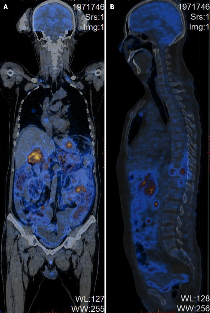
Case summary: A 53-year-old man was referred to our hospital from the local county hospital due to abdominal pain for two weeks and weakness in the lower extremities for one day. Magnetic resonance imaging of the abdomen and lumbar spine showed a swollen pancreas and gallbladder, with peripancreatic exudation and liquid collection, indicating acute pancreatitis and acute cholecystitis. Additionally, we observed abnormally thickened lesions of the gastric wall, multiple enlarged retroperitoneal lymph nodes and a well-demarcated, posterolateral extradural mass lesion between T9 and T12, with extension through the spinal foramen and definite bony destruction, suggesting metastasis in gastric malignancy. Subsequent whole-body positron emission tomography/computed tomography examination showed multifocal malignant lesions in the stomach, pancreas, gallbladder, bone, bilateral supraclavicular fossa, anterior mediastinum, bilateral axillary and retroperitoneal lymph nodes. Gastroduodenal endoscopy revealed primary BL with massive involvement of the gastric body and duodenum. The patient refused chemotherapeutic treatment and died one week later due to upper gastrointestinal hemorrhage. Afterward, we reviewed the characteristics of 11 patients with BL involving the stomach, pancreas or spinal cord.
Conclusion: Clinicians should be aware that BL can be the potential cause of acute pancreatitis or a rapidly progressive spinal tumor with accompanying paraplegia. For gastric BL, gastroscopy biopsies and pathology are necessary for a definite diagnosis.
Keywords: Acute pancreatitis; Burkitt lymphoma; Case report; Paraplegia.
©The Author(s) 2021. Published by Baishideng Publishing Group Inc. All rights reserved.
Conflict of interest statement
Conflict-of-interest statement: The authors declare that they have no conflicts of interest.
Figures


References
-
- Burkitt’s Lymphoma: Thorax to Pelvis. Indian J Chest Dis Allied Sci. 2016;58:49–51. - PubMed
-
- Goldman S, Smith L, Galardy P, Perkins SL, Frazer JK, Sanger W, Anderson JR, Gross TG, Weinstein H, Harrison L, Shiramizu B, Barth M, Cairo MS. Rituximab with chemotherapy in children and adolescents with central nervous system and/or bone marrow-positive Burkitt lymphoma/leukaemia: a Children's Oncology Group Report. Br J Haematol. 2014;167:394–401. - PMC - PubMed
-
- Dunleavy K, Little RF, Wilson WH. Update on Burkitt Lymphoma. Hematol Oncol Clin North Am. 2016;30:1333–1343. - PubMed
-
- Kelly JL, Toothaker SR, Ciminello L, Hoelzer D, Holte H, LaCasce AS, Mead G, Thomas D, Van Imhoff GW, Kahl BS, Cheson BD, Magrath IT, Fisher RI, Friedberg JW. Outcomes of patients with Burkitt lymphoma older than age 40 treated with intensive chemotherapeutic regimens. Clin Lymphoma Myeloma. 2009;9:307–310. - PMC - PubMed
Publication types
MeSH terms
LinkOut - more resources
Full Text Sources
Medical
