. 2021 Dec 22;7(12):2049-2062.
doi: 10.1021/acscentsci.1c01143. Epub 2021 Dec 8.

Ultrasound-Controlled CRISPR/Cas9 System Augments Sonodynamic Therapy of Hepatocellular Carcinoma

Affiliations

Ultrasound-Controlled CRISPR/Cas9 System Augments Sonodynamic Therapy of Hepatocellular Carcinoma

Haohao Yin et al. ACS Cent Sci. .

Abstract

Sonodynamic therapy (SDT), relying on the generation of reactive oxygen species (ROS), is a promising clinical therapeutic modality for the treatment of hepatocellular carcinoma (HCC) due to its noninvasiveness and high tissue-penetration depth, whereas the oxidative stress and antioxidative defense system in cancer cells significantly restrict the prevalence of SDT. Herein, we initially identified that NFE2L2 was immediately activated during SDT, which further inhibited SDT efficacy. To address this intractable issue, an ultrasound remote control of the cluster regularly interspaced short palindromic repeat (CRISPR)/CRISPR-associated protein 9 (Cas9) release system (HMME@Lip-Cas9) was meticulously designed and constructed, which precisely knocks down NFE2L2 to alleviate the adverse effects and augment the therapeutic efficiency of SDT. The hematoporphyrin monomethyl ether (HMME) in this system yielded abundant ROS to damage cancer cells under ultrasound irradiation, and meanwhile the generated ROS could induce lysosomal rupture to release Cas9/single guide RNA ribonucleoprotein (RNP) and destroy the oxidative stress-defensing system, significantly promoting tumor cell apoptosis. This study provides a new paradigm for HCC management and lays the foundation for the widespread application of CRISPR/Cas9 with promising clinical translation, meanwhile developing a synergistic therapeutic modality in the combination of SDT with gene editing.


Conflict of interest statement

The authors declare no competing financial interest.

Figures
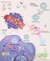
Figure 1
Figure 1
Schematic illustration of the designed strategy of the US-mediated CRISPR/Cas9 delivery system to enhance tumor SDT performance by amplifying oxidative stress. Preparation of HMME@Lip-Cas9 nanosystem and US-controlled CRISPR/Cas9 knock down target genes (NFE2L2). The CRISPR/Cas9 nanosystem (HMME@Lip-Cas9) generates ROS to promote apoptosis under ultrasound stimulation while disrupting the membrane structure of lysosomes, thus achieving the effective escape of Cas9/sgRNA RNP from lysosomes and efficient knock down of NFE2L2 for improving the therapeutic efficacy of SDT.
Figure 2
Figure 2
NFE2L2 promotes proliferation of HCC cells. (a) NFE2L2 protein expression levels after different SDT treatments by the Western blotting. (b) Immunofluorescence images of NFE2L2 (red) and nuclei stained by DAPI (blue) in tumors after different treatments. Scale bars, 50 μm. (c) Tumor growth curves (n = 5, mean ± SD) of hep3B2.1-7 cells with stable knock down of NFE2L2 in a xenograft mouse model (*p < 0.05, **p < 0.01, and ***p < 0.001). (d) Statistical and (e) photographic results of tumor size (n = 5, mean ± SD) in hep3B2.1-7 cells with stable knock down of NFE2L2 (*p < 0.05, **p < 0.01, and ***p < 0.001). (f) Tumor growth curves (n = 5, mean ± SD) of hep3B2.1-7 cells with stable overexpression of NFE2L2 in a xenograft mouse model (*p < 0.05, **p < 0.01, and ***p < 0.001). (g) Statistical and (h) photographic results of tumor size (n = 5, mean ± SD) in hep3B2.1-7 cells with stable overexpression of NFE2L2 (*p < 0.05, **p < 0.01, and ***p < 0.001). (i–j) Representative immunohistochemical images of Ki67 and PCNA protein levels in (i) hep3B2.1-7 xenografts with stable knock down of NFE2L2, and (j) hep3B2.1-7 xenografts with stable overexpression of NFE2L2, scale bars, 100 μm.
Figure 3
Figure 3
Characterizations of the HMME@Lip-Cas9 nanosystem. (a) Schematic illustration of the preparation and microstructure of HMME@Lip-Cas9 composite nanoparticles. (b) Agarose gel electrophoresis of HMME@Lip-Cas9 nanoparticles at different HMME@Lip/sgRNA ratios after incubation with serum (10% volume) for 6 h. (c) TEM image of HMME@Lip nanoparticles (scale bars: 200 nm). (d) TEM image of HMME@Lip-Cas9 composite nanoparticles (scale bars: 200 nm). (e) Zeta potential of Cas9/sgRNA, Lip, HMME@Lip, and HMME@Lip-Cas9 nanoparticles. (f) Hydrodynamic size distribution of Lip, HMME@Lip, and HMME@Lip-Cas9 nanoparticles. (g) Time-dependent UV–vis absorbance spectrum of DPBF under US irradiation. (h) Time-dependent UV–vis absorbance spectrum of DPBF treated with HMME@Lip-Cas9 nanoparticles under US irradiation. (i) UV–vis absorption values of DPBF at 410 nm after different treatments, including H2O + US and HMME@Lip-Cas9 + US. (j) ESR spectra of HMME@Lip-Cas9 + US + TEMP, and HMME@Lip-Cas9 + TEMP. (k) CLSM images of HepG2 cells stained with DCFH-DA after different treatments including control (without any treatment), US only, Cas9/sgRNA, HMME@Lip-Cas9, HMME@Lip + US (1.0 W cm–2), and HMME@Lip-Cas9 + US (1.0 W cm–2) (scale bars: 20 μm).
Figure 4
Figure 4
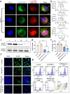
US-triggered HMME@Lip-Cas9 nanosystem endo-/lysosomal escape and US remote control of target gene knock down. (a) CLSM images and (b) corresponding mean fluorescence signal intensity of HepG2 cells cultured with Cy3-labeled HMME@Lip-Cas9 nanosystem (under US irradiation) for 1, 3, and 6 h at 37 °C. The cell nuclei were stained with DAPI (blue), endo-/lysosomes were stained with LysoTracker Green (green), and Cas9/sgRNA RNP was labeled with Cy3 (red). Scale bars, 10 μm. (c, d) The expression levels of EGFP in HepG2-EGFP cells after different treatments (I, control; II, Cas9/sgRNA; III, HMME@Lip-Cas9 without US; or IV, HMME@Lip-Cas9 with US) were analyzed by (c) Western blotting pictures and (d) semiquantitative analysis. (e, f) Typical CLSM images and corresponding average signal values of the fluorescence in different treated HepG2-EGFP cells (I, control; II, Cas9/sgRNA; III, HMME@Lip-Cas9 without US; or IV, HMME@Lip-Cas9 with US). Green, EGFP; blue, nuclei stained with DAPI. Scale bar, 20 μm. (g) FCM results reveal the gene editing efficiency of HepG2-EGFP cells after different treatments (I, control; II, Cas9/sgRNA; III, HMME@Lip-Cas9 without US; IV, HMME@Lip with US, or V, HMME@Lip-Cas9 with US). The EGFP-silencing efficiency of HMME@Lip-Cas9 and HMME@Lip-Cas9 was 44.2% and 78.2%, respectively.
Figure 5
Figure 5
Synergistic therapeutic effects of gene editing and sonodynamic therapy. (a) Schematic illustration of the gene locus of the target gene NFE2L2. (b) CCK8 results showing the viability of HepG2 cells after coculture with different concentrations of the HMME@Lip-Cas9 nanosystem (0, 25, 50, 100, 200, and 400 μg mL–1) for 24 h (n = 6, *p < 0.05, **p < 0.01 and ***p < 0.001). (c) Images of clone formation and (d) the corresponding clone formation rate of HepG2 cells after different treatments, including control, US only, Cas9/sgRNA, HMME@Lip-Cas9, HMME@Lip + US, or HMME@Lip-Cas9 + US (n = 3, *p < 0.05, **p < 0.01 and ***p < 0.001). (e) CCK-8 assay of HepG2 cells after different treatments, including control, US only, Cas9/sgRNA, HMME@Lip-Cas9, HMME@Lip + US, and HMME@Lip-Cas9 + US (n = 4, *p < 0.05, **p < 0.01, and ***p < 0.001). (f) CLSM images of HepG2 cells stained by calcein-AM (green) and PI (red) after different treatments, including control, US only, Cas9/sgRNA, HMME@Lip-Cas9, HMME@Lip + US, or HMME@Lip-Cas9 + US. Scale bar: 50 μm. (g) DNA sequencing results of NFE2L2 reveal the knock down capability of HepG2 cells after different treatments (control, US only, Cas9/sgRNA, HMME@Lip-Cas9, HMME@Lip + US, or HMME@Lip-Cas9 + US). (h, i) NGS results showing indel percentage of NFE2L2 in HepG2 cells treated with (h) HMME@Lip-Cas9 and (i) HMME@Lip-Cas9 + US.
Figure 6
Figure 6
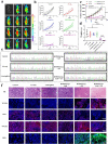
NFE2L2 gene editing therapy/SDT-based synergistic therapy in vivo. (a) In vivo fluorescence imaging of mice with HepG2 tumors at various time points after tail vein injection of the HMME@Lip-Cas9 nanosystem (0, 0.5, 1, 2, 3, 4, 6, 8, 12, 24, 36, and 48 h). (b) Tumor growth curves of HepG2 tumor-bearing mice after different treatments (control, US only, Cas9/sgRNA, HMME@Lip-Cas9, HMME@Lip + US, and HMME@Lip-Cas9 + US). (c) Time-dependent tumor growth curves (n = 5, mean ± SD) after various treatments (*p < 0.05, **p < 0.01, and ***p < 0.001). (d) Tumor weight after different treatments. (e) Sanger sequencing of NFE2L2 in HepG2 tumor-bearing mice after 24 h of exposure to varying treatments. (f) HepG2 tumor-bearing mice underwent HE staining and immunofluorescence staining (TUNEL, Ki67, NFE2L2, DCFH-DA) after 24 h of different treatments. Nuclei were stained by DAPI (blue).
Figure 7
Figure 7
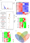
Mechanism of synergistic treatment of HMME@Lip-Cas9 under US irradiation. (a) Volcano map of genetic alterations after HMME@Lip-Cas9 treatment compared to control (p < 0.05, |fold change| ≥ 2). (b) Heat map of genetic alterations after HMME@Lip-Cas9 treatment compared to control (p < 0.05, |fold change| ≥ 2). (c) KEGG analysis of differential gene expression profiles based on the results of transcriptome sequencing after synergistic gene-editing therapy/SDT of tumors. (d) Heatmap of genes altering in protein processing in the p53 signaling pathway. (e) Heatmap of genes altering in protein processing in the apoptosis signaling pathway. (f) Venn diagram of the differentially expressed genes (DEGs) involved in cell cycle, MAPK, p53, and apoptotic signaling pathways.

References

    1. Tang A.; Hallouch O.; Chernyak V.; Kamaya A.; Sirlin C. B. Epidemiology of hepatocellular carcinoma: target population for surveillance and diagnosis. Abdom. Radiol. (NY) 2018, 43 (1), 13–25. 10.1007/s00261-017-1209-1. - DOI - PubMed
    1. Sung H.; Ferlay J.; Siegel R. L.; Laversanne M.; Soerjomataram I.; Jemal A.; Bray F. Global cancer statistics 2020: GLOBOCAN estimates of incidence and mortality worldwide for 36 cancers in 185 countries. Ca-Cancer J. Clin. 2021, 71 (3), 209–249. 10.3322/caac.21660. - DOI - PubMed
    1. Cabibbo G.; Enea M.; Attanasio M.; Bruix J.; Craxi A.; Camma C. A meta-analysis of survival rates of untreated patients in randomized clinical trials of hepatocellular carcinoma. Hepatology 2010, 51 (4), 1274–83. 10.1002/hep.23485. - DOI - PubMed
    1. Bruix J.; Takayama T.; Mazzaferro V.; Chau G. Y.; Yang J.; Kudo M.; Cai J.; Poon R. T.; Han K. H.; Tak W. Y.; et al. Adjuvant sorafenib for hepatocellular carcinoma after resection or ablation (STORM): a phase 3, randomised, double-blind, placebo-controlled trial. Lancet Oncol. 2015, 16 (13), 1344–54. 10.1016/S1470-2045(15)00198-9. - DOI - PubMed
    1. Guan W.; Tan L.; Liu X.; Cui Z.; Zheng Y.; Yeung K. W. K.; Zheng D.; Liang Y.; Li Z.; Zhu S.; Wang X.; Wu S. Ultrasonic Interfacial Engineering of Red Phosphorous-Metal for Eradicating MRSA Infection Effectively. Adv. Mater. 2021, 33 (5), e200604710.1002/adma.202006047. - DOI - PubMed