. 2022 Jan 15;208(2):407-419.
doi: 10.4049/jimmunol.2100029. Epub 2021 Dec 29.

Mucosal Vaccination with Cyclic Dinucleotide Adjuvants Induces Effective T Cell Homing and IL-17-Dependent Protection against Mycobacterium tuberculosis Infection

Affiliations

Mucosal Vaccination with Cyclic Dinucleotide Adjuvants Induces Effective T Cell Homing and IL-17-Dependent Protection against Mycobacterium tuberculosis Infection

Robyn M Jong et al. J Immunol. .

Abstract

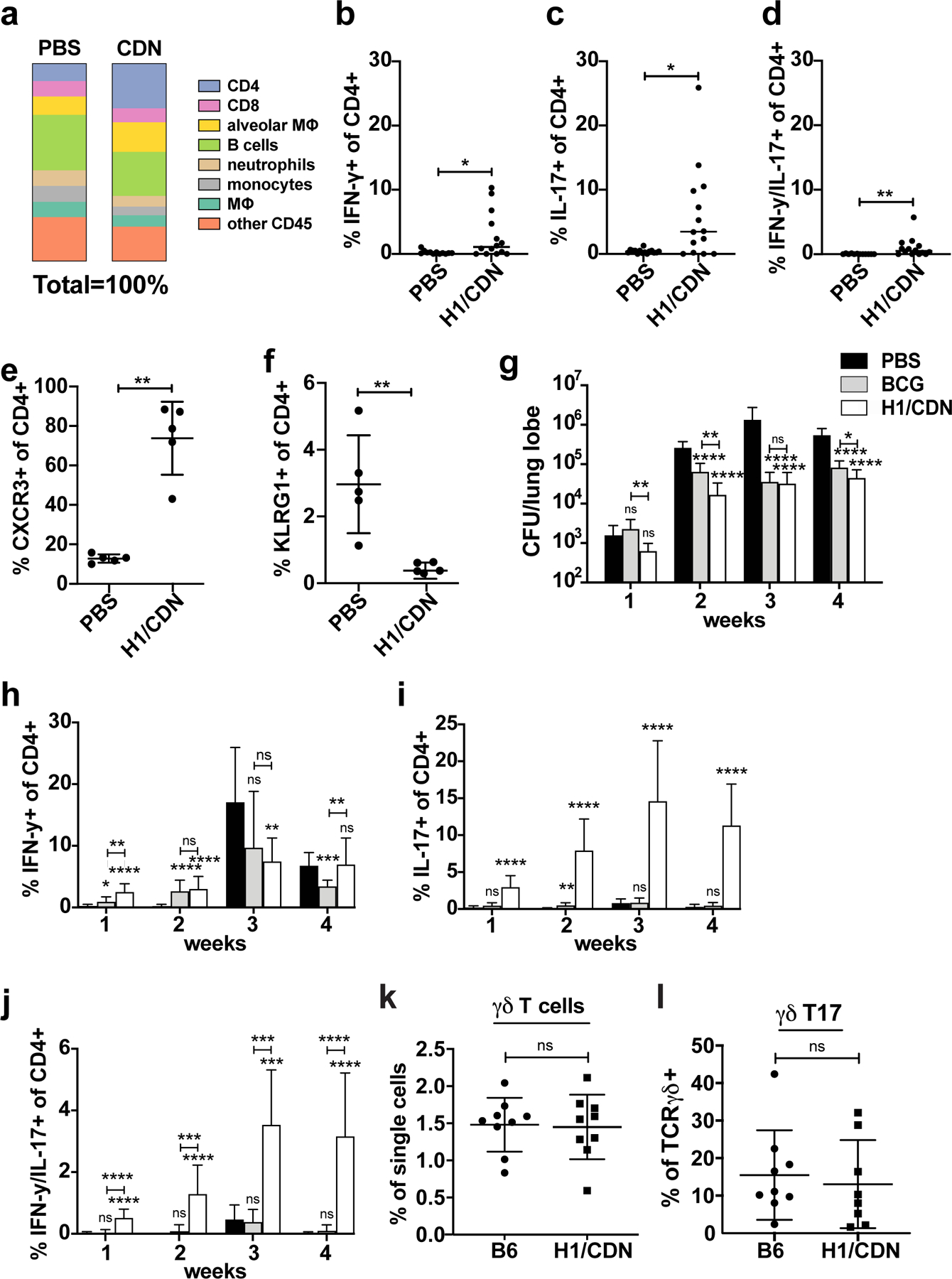
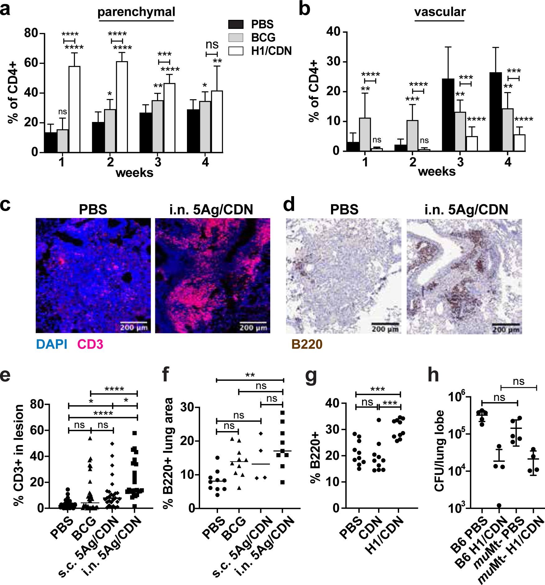
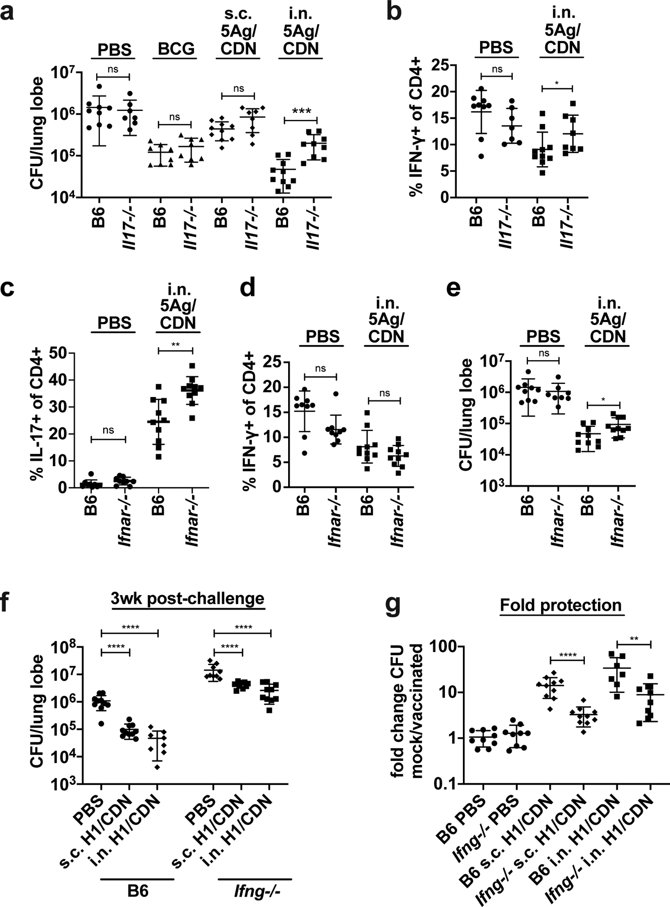
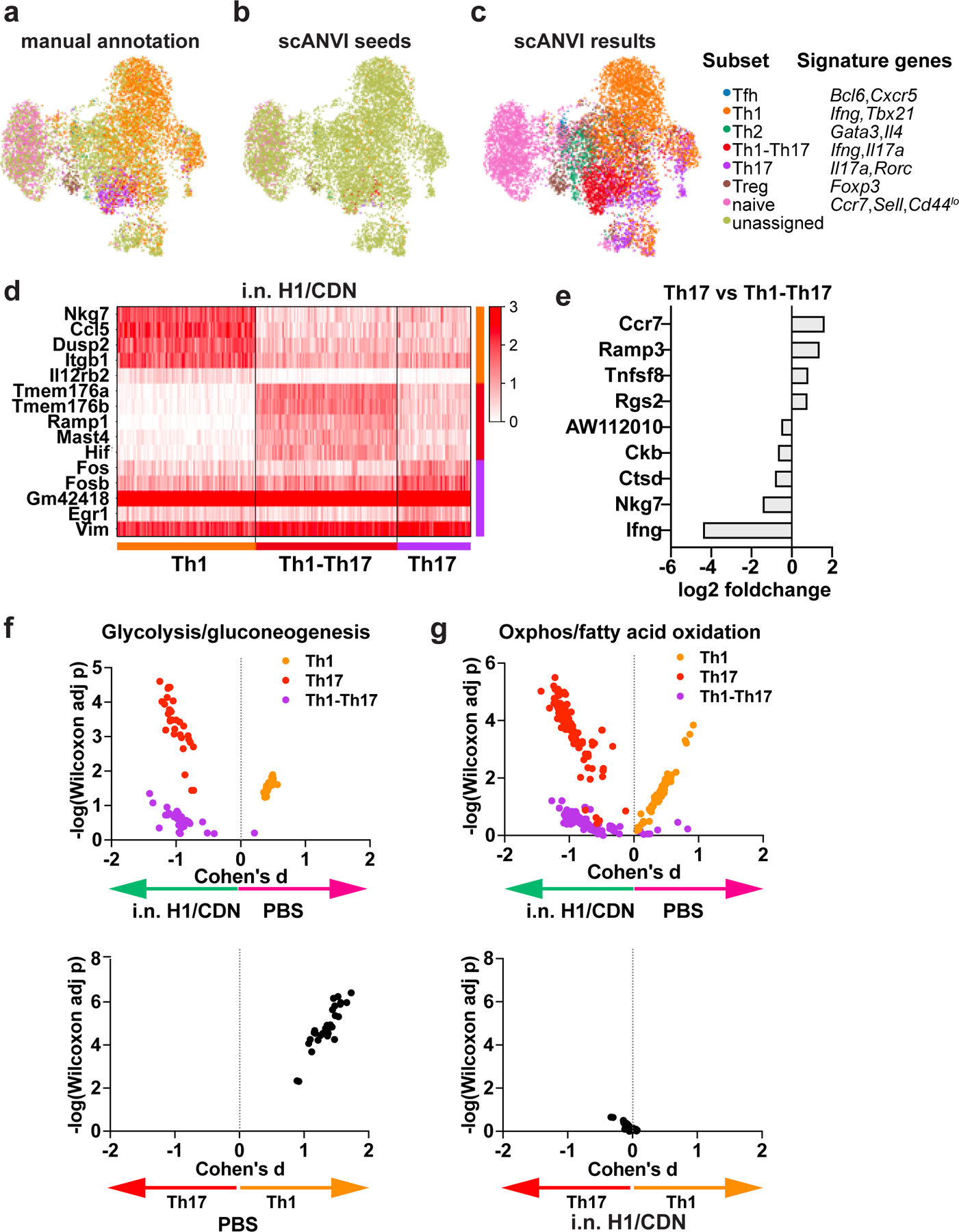
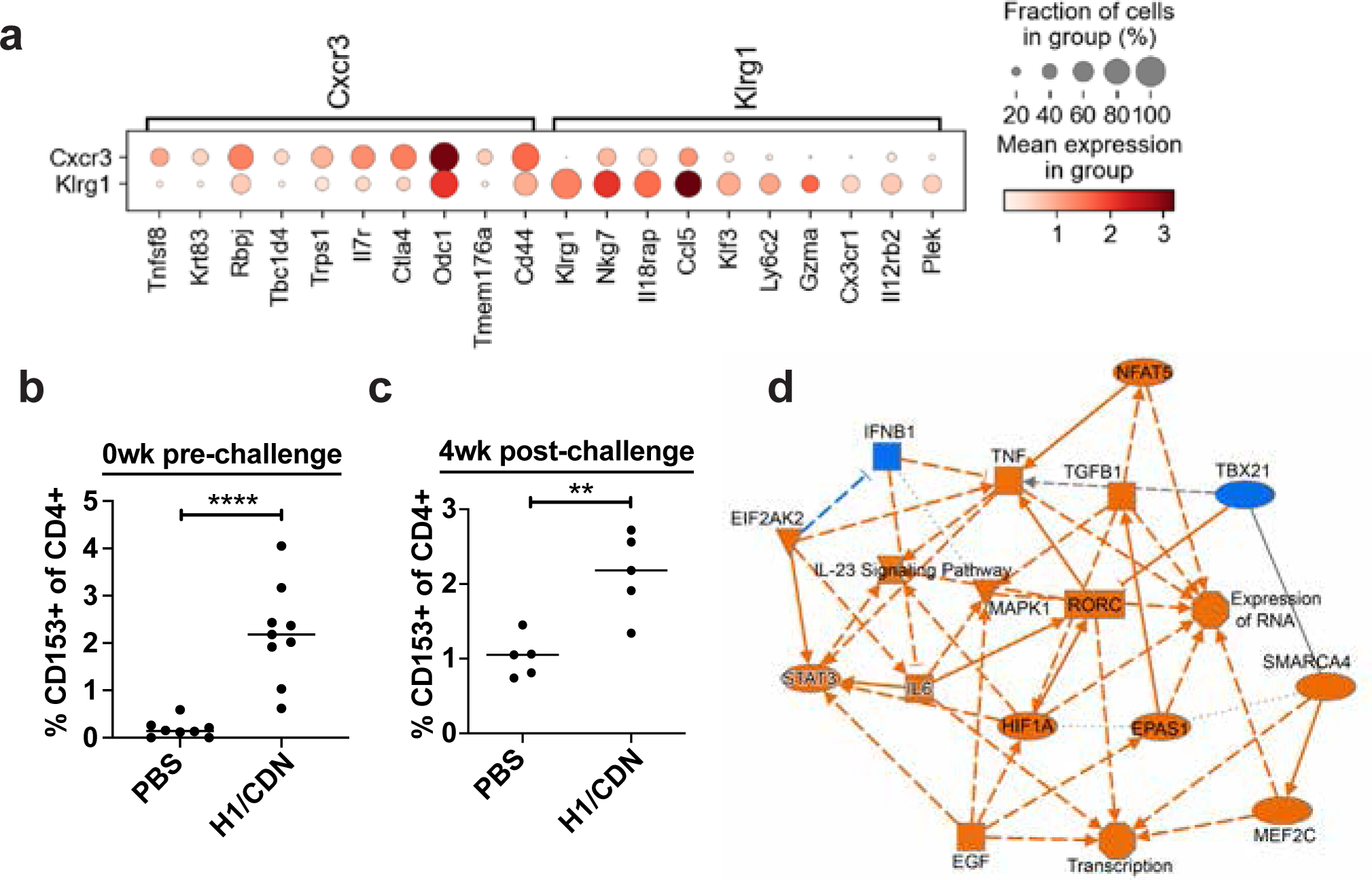
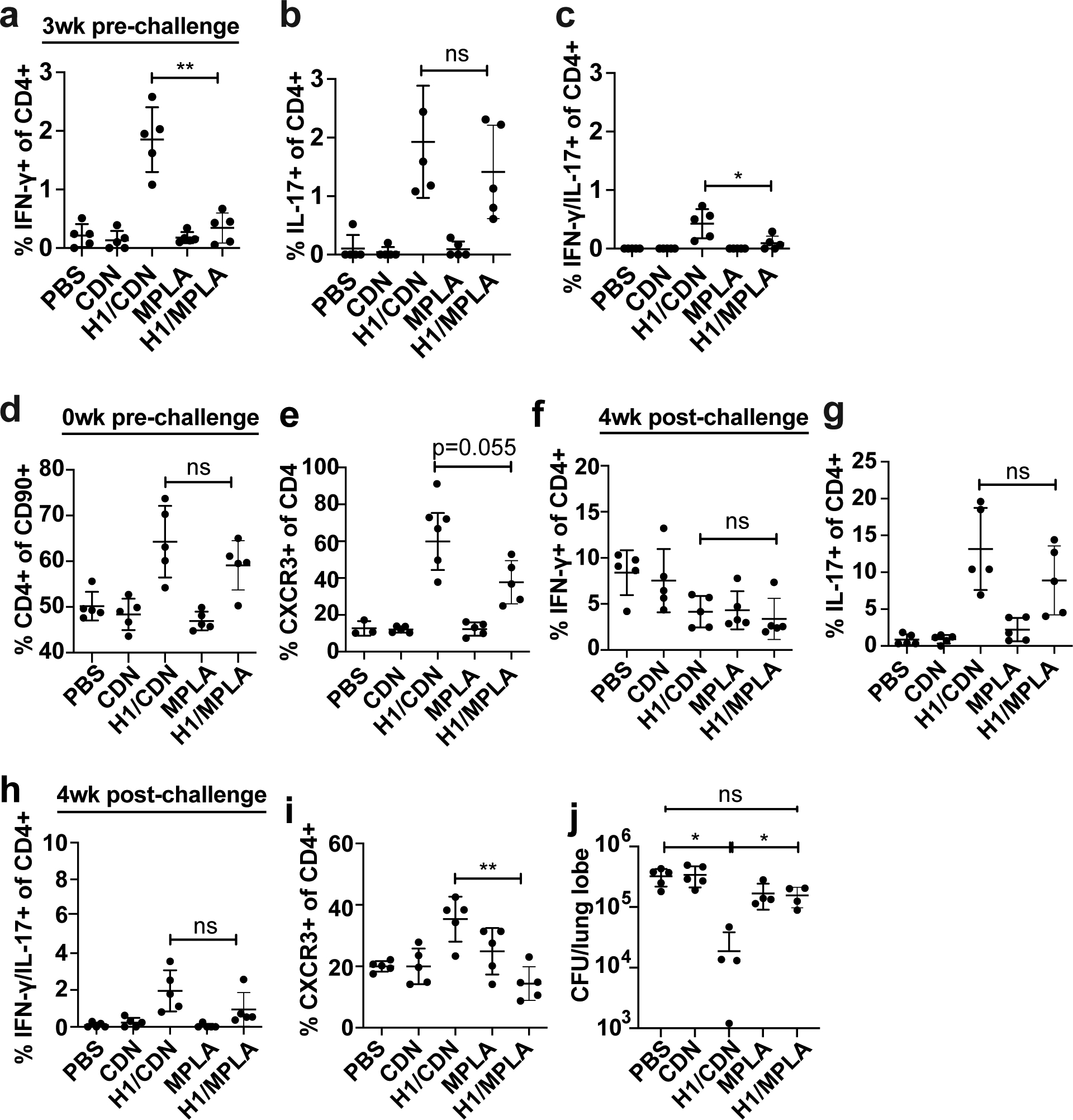
Tuberculosis consistently causes more deaths worldwide annually than any other single pathogen, making new effective vaccines an urgent priority for global public health. Among potential adjuvants, STING-activating cyclic dinucleotides (CDNs) uniquely stimulate a cytosolic sensing pathway activated only by pathogens. Recently, we demonstrated that a CDN-adjuvanted protein subunit vaccine robustly protects against tuberculosis infection in mice. In this study, we delineate the mechanistic basis underlying the efficacy of CDN vaccines for tuberculosis. CDN vaccines elicit CD4 T cells that home to lung parenchyma and penetrate into macrophage lesions in the lung. Although CDNs, like other mucosal vaccines, generate B cell-containing lymphoid structures in the lungs, protection is independent of B cells. Mucosal vaccination with a CDN vaccine induces Th1, Th17, and Th1-Th17 cells, and protection is dependent upon both IL-17 and IFN-γ. Single-cell RNA sequencing experiments reveal that vaccination enhances a metabolic state in Th17 cells reflective of activated effector function and implicate expression of Tnfsf8 (CD153) in vaccine-induced protection. Finally, we demonstrate that simply eliciting Th17 cells via mucosal vaccination with any adjuvant is not sufficient for protection. A vaccine adjuvanted with deacylated monophosphoryl lipid A (MPLA) failed to protect against tuberculosis infection when delivered mucosally, despite eliciting Th17 cells, highlighting the unique promise of CDNs as adjuvants for tuberculosis vaccines.


Figures

Figure 1.
Figure 1.. Intranasal immunization with H1/CDN enhances CD4 T cell responses in the lungs of vaccinated mice both pre and post-challenge.
Mice were vaccinated with intranasal M. tuberculosis H1 antigen and ML-RR-cGAMP cyclic-di-nucleotide adjuvant (H1/CDN) 12 weeks before challenge, then boosted twice at 4-week intervals after priming, before M. tuberculosis aerosol challenge with 50–100 CFU. Mice were mock primed and boosted with PBS as a control. (a) Cellular percentages were enumerated in the lungs using flow cytometry in vaccinated and mock vaccinated mice on day 0 prior to challenge with M. tuberculosis. (b) Intracellular cytokine staining (ICS) for percentage of lung CD4 T cells that produce IFN-γ (Th1), (c) IL-17 (Th17), (d) both IFN-γ and IL-17 (Th1-Th17) after restimulation ex vivo with recombinant Ag85b peptide pool on day 0 prior to challenge with M. tuberculosis. Percentage of CD4 T cells that express (e) CXCR3 (parenchymal T cells) or (f) KLRG1 (vascular T cells) on day 0 prior to challenge with M. tuberculosis. Mice were vaccinated with s.c. BCG or i.n. H1/CDN as above and (g) lung bacterial burden was enumerated at 1 – 4 weeks post M. tuberculosis challenge. (h) Intracellular cytokine staining (ICS) for percentage of lung CD4 T cells that produce IFN-γ (Th1) (i) IL-17 (Th17), or (j) both IFN-γ and IL-17 (Th1-Th17) after restimulation ex vivo with recombinant Ag85b peptide pool at indicated timepoints post challenge. (k) Enumeration of TCRɣδ and (l) IL-17-producing TCRɣδ T cells at 4 weeks post challenge. Data are expressed as mean (± SD) of eight to ten mice per group from two independent experiments; in (b-f; k, l) each animal is represented by a point. Mann-Whitney t test p values calculated in comparison to PBS vaccinated controls except where indicated; *p ≤ 0.05, **p ≤ 0.01, ***p ≤ 0.001, ****p ≤ 0.0001. Significance is relative to PBS control unless otherwise indicated. All experiments were performed a minimum of two times; for a-d two pooled experiments are shown; otherwise a representative experiment is shown.
Figure 2.
Figure 2.. Intranasal immunization with H1/CDN promotes T cell influx into macrophage aggregates in lungs.
Mice were immunized as described and analyzed at indicated timepoints. (a) Surface staining for percentage of parenchymal CXCR3+ KLRG1 lung CD4 T cells from mock and i.n. H1/ML-RR-cGAMP (i.n. H1/CDN) vaccinated mice. (b) Surface staining for percentage of vascular CXCR3 KLRG1+ lung CD4 T cells. Mice were immunized as described and analyzed at 4 weeks post challenge with M. tuberculosis. (c) Representative immunofluorescent staining of formalin-fixed, paraffin-embedded lung sections from mock and 5Ag/ML-RR-cGAMP-vaccinated mice for T cell marker CD3 and nuclear DAPI stain. (d) Representative immunohistochemical staining of lung sections for B cell marker B220. (e) Quantification of CD3+ immune cells out of total cells in lung lesions. (f) Quantification of B220+ lung area. (g) Percentage of B220+ cells in the lungs at 4 weeks post challenge analyzed by flow cytometry. (h) WT and muMT mice were immunized with H1/CDN and M. tuberculosis CFU in the lungs was enumerated at 4 weeks post challenge. Data are expressed as mean (± SD) of eight to ten mice per group from two independent experiments; each animal is shown as an individual point. (a, b, g, h) Mann-Whitney t test p values; (e, f) Kruskal Wallis test followed by Dunn multiple comparison posthoc p-values; *p ≤ 0.05, **p ≤ 0.01, ***p ≤ 0.001, ****p ≤ 0.0001. Significance is relative to PBS control unless otherwise indicated. All experiments were performed a minimum of twice; a-f a representative experiment is shown, for g-h pooled data from 2 experiments is shown.
Figure 3.
Figure 3.. IL-17, Type I IFN, and IFN-γ all contribute to vaccine-elicited control of infection with M. tuberculosis.
Wild-type C57BL/6 (B6) and Il17−/− mice were vaccinated once with subcutaneous M. bovis BCG 12 weeks before M. tuberculosis aerosol challenge or primed with either subcutaneous or intranasal M. tuberculosis 5Ag antigen and ML-RR-cGAMP cyclic-di-nucleotide adjuvant (i.n. 5Ag/CDN) 12 weeks before challenge, followed by two boosts. (a) At 4 weeks post challenge, mouse lungs were harvested and analyzed for bacterial burden. (b) ICS analysis of ex vivo restimulated lung leukocytes for IFN-γ. (c) Wild-type C57BL/6 (B6) and Ifnar−/− mice were vaccinated as described and CD4 T cells expressing IL-17 or (d) IFN-γ were evaluated by ICS and flow cytometry at 4 weeks post challenge. (e) Bacterial burdens in the lungs were enumerated at 4 weeks post challenge. Wild-type C57BL/6 (B6) and Ifng−/− mice were vaccinated as described and (f) bacterial burden was enumerated at 4 weeks post challenge. (g) data is reiterated in (f) expressed as fold change. Data are expressed as mean (± SD) of eight to ten mice per group from two independent experiments pooled with value from each individual animal displayed. Mann-Whitney t test p values; *p ≤ 0.05, **p ≤ 0.01, ***p ≤ 0.001.
Figure 4.
Figure 4.. scRNA-seq analysis of CD4 T cells reveals vaccine-elicited differences in CD4 T cells subsets and shifts in Th17 metabolism.
Subsets of CD4 T cells were (a) manually annotated based on signature expression of lineage-specific transcription factor, cytokine, and cell surface markers or (b, c) annotated using scANVI. (d) Heatmap depicting genes that were highly expressed in Th1, Th1-Th17, and Th17 cells. (e) Genes that were differentially expressed in Th17 vs Th1-Th17 cells. (f) Compass analysis of glycolysis in Th1, Th17, and Th1-Th17 subsets. (g) Compass analysis of oxidative phosphorylation in Th1, Th17, and Th1-Th17 subsets.
Figure 5.
Figure 5.. Vaccination enhances expression of Tnfsf8 (CD153) in parenchyma-homing T cells and in the CD4 T cell compartment.
Mice were vaccinated three times at 4 week intervals with intranasal H1/CDN. (a) scRNA-seq results from all CXCR3+KLRG1 vs. CXCR3KLRG1+ CD4 T cells were compared. Top genes that were differentially expressed in these subtypes. (b) Flow cytometry analysis for CD153 expression on CD4 T cells at day 0 prior to challenge or (c) at 4 weeks post challenge. (d) Network analysis of scRNA-seq results comparing CXCR3+KLRG1 and CXCR3KLRG1+. Orange nodes are overrepresented in CXCR3+ cells; blue nodes are overrepresented in KLRG1+ cells.
Figure 6.
Figure 6.. Vaccination with H1/CDN by the intranasal route elicits more robust protection than vaccination with H1/MPLA.
Mice were vaccinated three times at 4 week intervals with either intranasal H1/CDN or H1/MPLA. (a) Intracellular cytokine staining (ICS) for percentage of blood CD4 T cells that produce IFN-γ, (b) IL-17, or (c) IFN-γ and IL-17 at 3 weeks prior to challenge. (d) Flow cytometry analysis for CD4 expression on T cells or (e) CXCR3 expression on CD4 T cells at day 0 prior to challenge. (f) Intracellular cytokine staining (ICS) for percentage of lung CD4 T cells that produce IFN-γ, (g) IL-17, or (h) IFN-γ and IL-17 at 4 weeks after challenge with M. tuberculosis. (i) Flow cytometry analysis for CXCR3 expression on T cells at 4 weeks post challenge. (j) CFU from mice vaccinated with CDN or MPLA-adjuvanted vaccines at 4 weeks post challenge. Data are expressed as mean (± SD) of five mice per group with value from each individual animal displayed. Each experiment was performed a minimum of twice and a representative experiment is shown. Mann-Whitney t test p values; *p ≤ 0.05, **p ≤ 0.01, ***p ≤ 0.001.

Similar articles

Cited by

References

    1. Geneva. 2018. Global tuberculosis report 2018,. World Health Organization.
    1. Colditz GA 1994. The efficacy of bcg vaccine in the prevention of tuberculosis. Pediatr. Infect. Dis. J 13: 761.
    1. Fine PE 1995. Variation in protection by BCG: implications of and for heterologous immunity. Lancet 346: 1339–1345. - PubMed
    1. Mangtani P, Abubakar I, Ariti C, Beynon R, Pimpin L, Fine PEM, Rodrigues LC, Smith PG, Lipman M, Whiting PF, and Sterne JA. 2014. Protection by BCG vaccine against tuberculosis: a systematic review of randomized controlled trials. Clin. Infect. Dis 58: 470–480. - PubMed
    1. Cooper AM 2009. Cell-mediated immune responses in tuberculosis. Annu. Rev. Immunol 27: 393–422. - PMC - PubMed

Publication types

MeSH terms