. 2022 Feb 16;42(7):1352-1361.
doi: 10.1523/JNEUROSCI.1601-21.2021. Epub 2021 Dec 29.

Distinct Factors Drive the Spatiotemporal Progression of Tau Pathology in Older Adults

Affiliations

Distinct Factors Drive the Spatiotemporal Progression of Tau Pathology in Older Adults

Jenna N Adams et al. J Neurosci. .

Abstract

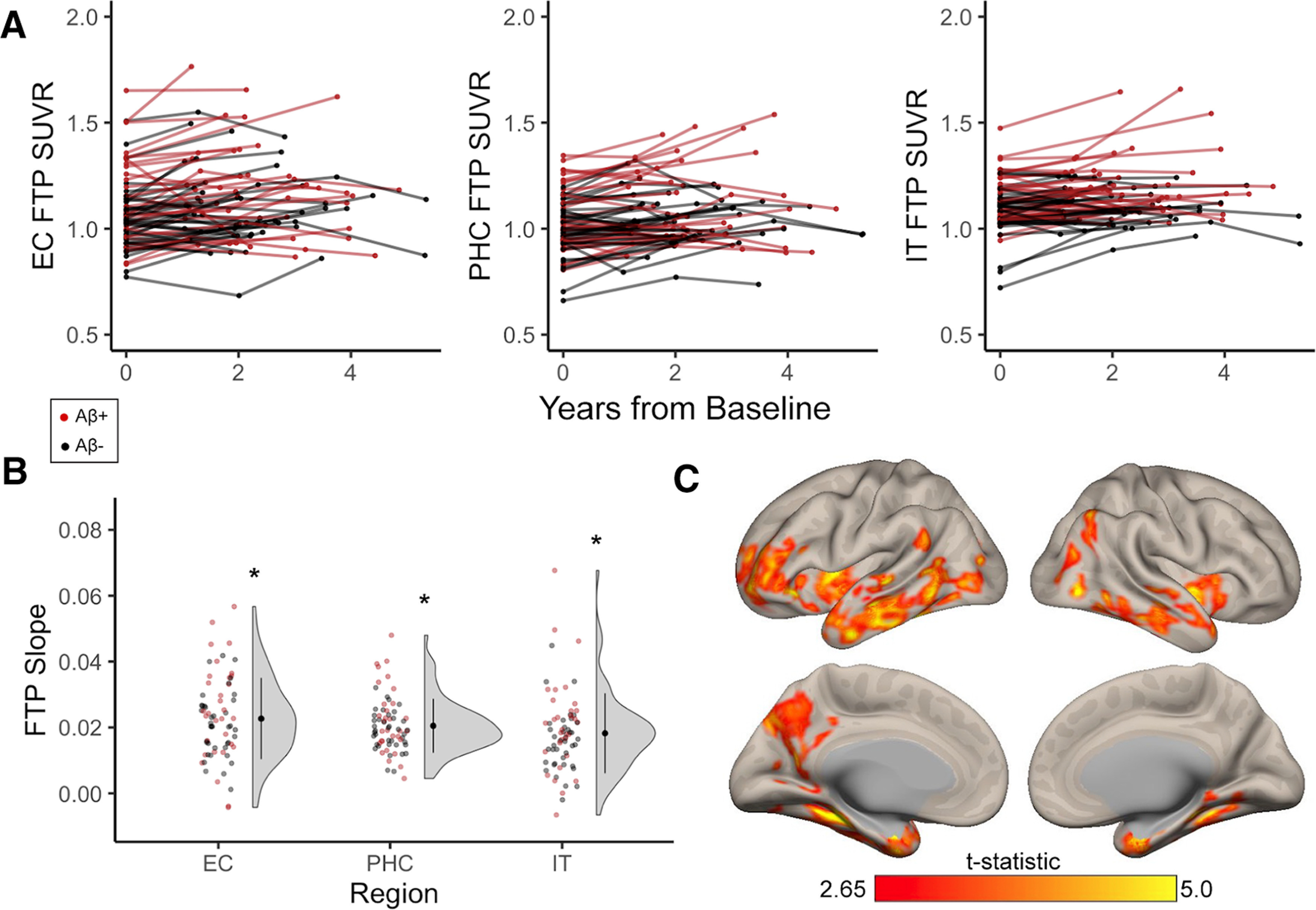
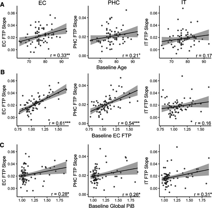
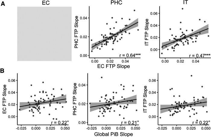
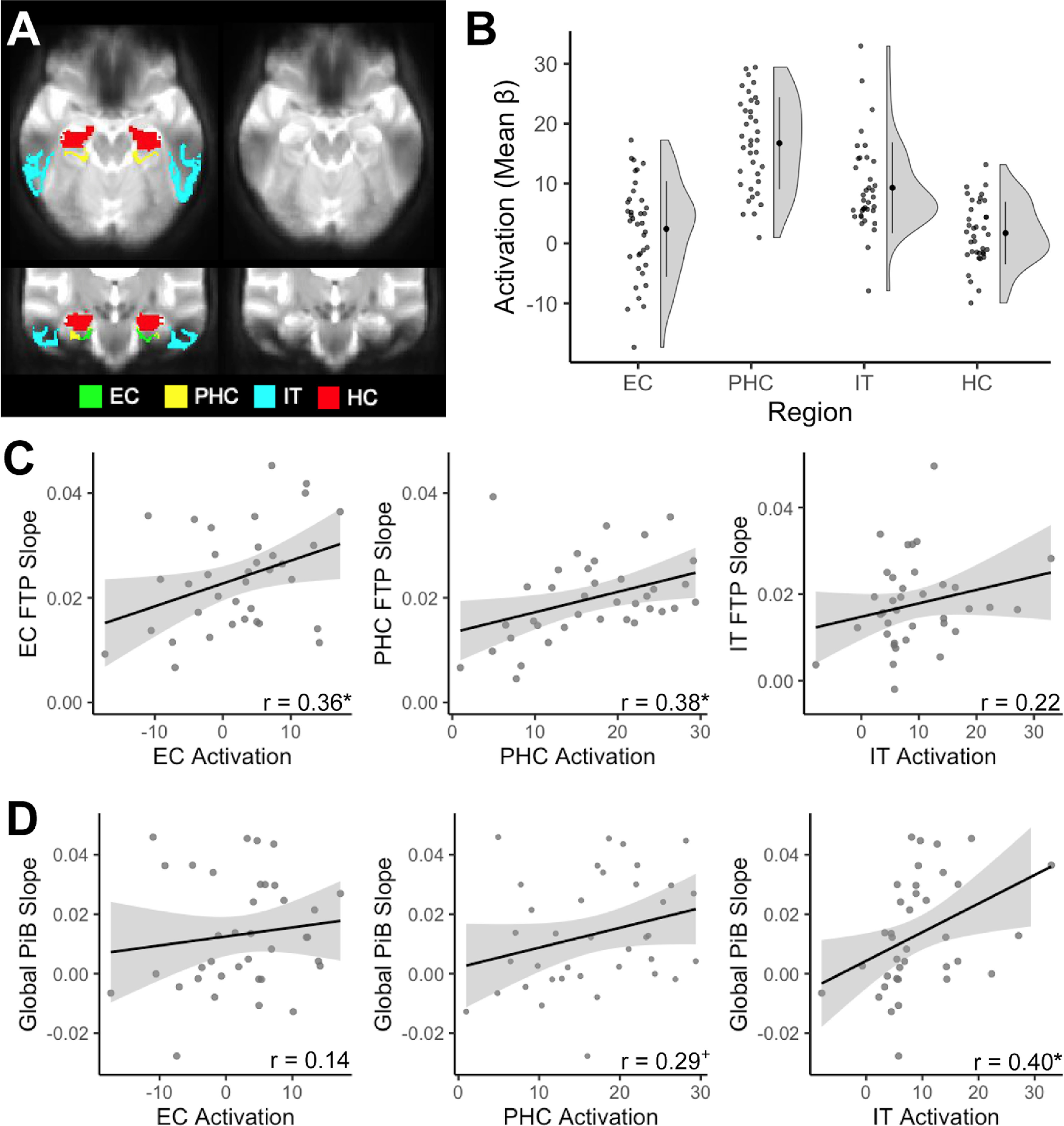
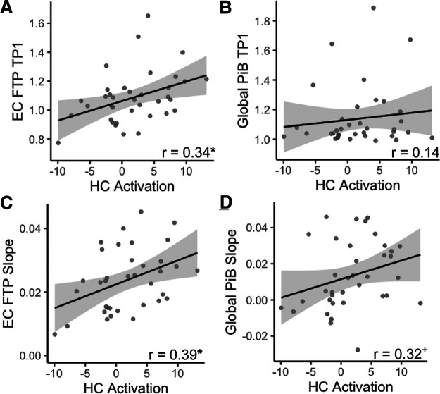
Mechanisms underlying the initial accumulation of tau pathology across the human brain are largely unknown. We examined whether baseline factors including age, amyloid-β (Aβ), and neural activity predicted longitudinal tau accumulation in temporal lobe regions that reflect distinct stages of tau pathogenesis. Seventy cognitively normal human older adults (77 ± 6 years, 59% female) received two or more 18F-flortaucipir (FTP) and 11C-Pittsburgh Compound B (PiB) PET scans (mean follow-up, 2.5 ± 1.1 years) to quantify tau and (Aβ). Linear mixed-effects models were used to calculate the slopes of FTP change in entorhinal cortex (EC), parahippocampal cortex (PHC), and inferior temporal gyrus (IT), and slopes of global PiB change. Thirty-seven participants underwent functional MRI to measure baseline activation. Older age predicted EC tau accumulation, and baseline EC tau levels predicted subsequent tau accumulation in EC and PHC. In IT, however, baseline EC tau interacted with Aβ to predict IT tau accumulation. Higher baseline local activation predicted tau accumulation within EC and PHC, and higher baseline hippocampal activation predicted EC tau accumulation. Our findings indicate that factors predicting tau accumulation vary as tau progresses through the temporal lobe. Older age is associated with initial tau accumulation in EC, while baseline EC tau and neural activity drive tau accumulation within medial temporal lobe. Aβ subsequently facilitates tau spread from medial to lateral temporal lobe. Our findings elucidate potential drivers of tau accumulation and spread in aging, which are critical for understanding Alzheimer's disease pathogenesis.SIGNIFICANCE STATEMENT To further understand the mechanisms leading to tau pathogenesis and spread, we tested whether baseline factors such as age, amyloid-β pathology, and activation predicted longitudinal tau accumulation in cognitively normal older adults. We found that distinct mechanisms contribute to tau accumulation as tau progresses across the temporal lobe, with initial tau accumulation in entorhinal cortex driven by age and subsequent spread driven by neural activity and amyloid-β. We demonstrate that higher baseline activation predicts increased longitudinal tau accumulation, providing novel evidence that activation-dependent tau production may occur in the human brain. Our findings support major hypotheses generated from preclinical research, and have important translational implications, suggesting that the reduction of hyperactivation may help prevent the development of tau pathology.

Keywords: Alzheimer's disease; PET; aging; amyloid-β; fMRI; tau.


Figures

Figure 1.
Figure 1.
Longitudinal tau accumulation measured with FTP in cognitively normal older adults. Cognitively normal older adults received at least two time points of FTP, which was processed with a longitudinal pipeline. A, B, Red data points indicate participants who were Aβ+ at baseline, while black data points indicate Aβ− participants. A, Individual trajectories of FTP SUVRs over longitudinal follow-up in entorhinal cortex (EC), parahippocampal cortex (PHC), and inferior temporal gyrus (IT). B, FTP slopes were computed to quantify longitudinal change in tau with linear mixed-effects models. FTP slopes were significantly greater than zero (one-sample t tests, *p < 0.001) in EC, PHC, and IT, indicating group-level tau accumulation in these regions. C, Whole-brain voxelwise FTP accumulation occurred predominantly in temporal regions (overlapping with a priori regions), but also in small clusters in medial parietal and frontal lobe, replicating previous findings in a subset of this sample. See Extended Data Figure 1-1 for whole-brain visualization of mean FTP slope in Aβ− and Aβ+ groups separately.
Figure 2.
Figure 2.
Baseline predictors of longitudinal tau accumulation. Pearson correlations between FTP slope, indicating the rate of longitudinal tau accumulation, and baseline risk factors. A, Higher age at baseline significantly predicted increased FTP slope in entorhinal cortex (EC). B, Higher baseline EC FTP was associated with increased FTP slope in EC and parahippocampal cortex (PHC), but not in inferior temporal gyrus (IT). C, Higher baseline global PiB was associated with increased FTP slope in EC, PHC, and IT. ***p < 0.001, **p < 0.01, *p < 0.05, +p < 0.10.
Figure 3.
Figure 3.
Longitudinal associations between tau and Aβ accumulation. A, FTP slope in entorhinal cortex (EC) was highly correlated with FTP slope in parahippocampal cortex (PHC) and inferior temporal gyurs (IT). Gray shading indicates that this analysis could not be performed due to the autocorrelation between EC FTP slope. B, Global PiB slope was not significantly associated with FTP slope in any region, though these associations were trending toward significance. ***p < 0.001, +p < 0.10.
Figure 4.
Figure 4.
Local activation during memory processing and longitudinal accumulation of Alzheimer's pathology. A, We investigated activity in entorhinal cortex (EC; green), parahippocampal cortex (PHC; yellow), inferior temporal gyrus (IT; cyan), and hippocampus (HC; red). Signal dropout predominantly occurred in lateral temporal regions, affecting IT; however, we removed low-signal voxels before analyses. B, Activation (mean β-values) was extracted for each region, contrasting all task images against the perceptual baseline to derive a general measure of activation during memory processing. C, Baseline activation was associated with increased FTP slope locally within EC and PHC, but not IT. D, Baseline IT activation, but not EC or PHC activation, was associated with increased global PiB slope. *p < 0.05, +p < 0.10.
Figure 5.
Figure 5.
Hippocampal (HC) activation and longitudinal accumulation of Alzheimer's pathology. A, B, At baseline, increased HC activation was significantly associated with FTP in entorhinal cortex (EC) (A), but not global PiB (B). C, D, Higher baseline HC activation predicted increased FTP slope in EC (C), and a trend-level association with increased global PiB slope (D). *p < 0.05, +p < 0.10.

References

    1. Adams JN, Maass A, Harrison TM, Baker SL, Jagust WJ (2019) Cortical tau deposition follows patterns of entorhinal functional connectivity in aging. Elife 8:e49132. 10.7554/eLife.49132 - DOI - PMC - PubMed
    1. Adams JN, Maass A, Berron D, Harrison TM, Baker SL, Thomas WP, Stanfill M, Jagust WJ (2021) Reduced repetition suppression in aging is driven by tau-related hyperactivity in medial temporal lobe. J Neurosci 41:3917–3931. 10.1523/JNEUROSCI.2504-20.2021 - DOI - PMC - PubMed
    1. Baker SL, Maass A, Jagust WJ (2017) Considerations and code for partial volume correcting [18F]-AV-1451 tau PET data. Data Brief 15:648–657. 10.1016/j.dib.2017.10.024 - DOI - PMC - PubMed
    1. Bakker A, Krauss GL, Albert MS, Speck CL, Jones LR, Stark CE, Yassa MA, Bassett SS, Shelton AL, Gallagher M (2012) Reduction of hippocampal hyperactivity improves cognition in amnestic mild cognitive impairment. Neuron 74:467–474. 10.1016/j.neuron.2012.03.023 - DOI - PMC - PubMed
    1. Berron D, Neumann K, Maass A, Schütze H, Fliessbach K, Kiven V, Jessen F, Sauvage M, Kumaran D, Düzel E (2018) Age-related functional changes in domain-specific medial temporal lobe pathways. Neurobiol Aging 65:86–97. 10.1016/j.neurobiolaging.2017.12.030 - DOI - PubMed

Publication types