. 2021 Dec 13:12:766821.
doi: 10.3389/fimmu.2021.766821. eCollection 2021.

A Potent and Protective Human Neutralizing Antibody Against SARS-CoV-2 Variants

Affiliations

A Potent and Protective Human Neutralizing Antibody Against SARS-CoV-2 Variants

Sisi Shan et al. Front Immunol. .

Abstract

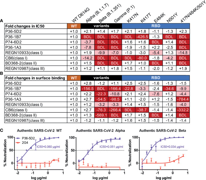
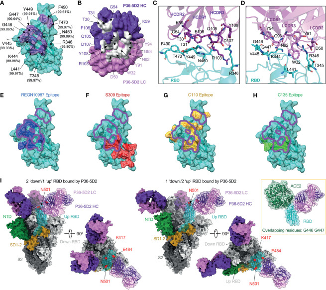
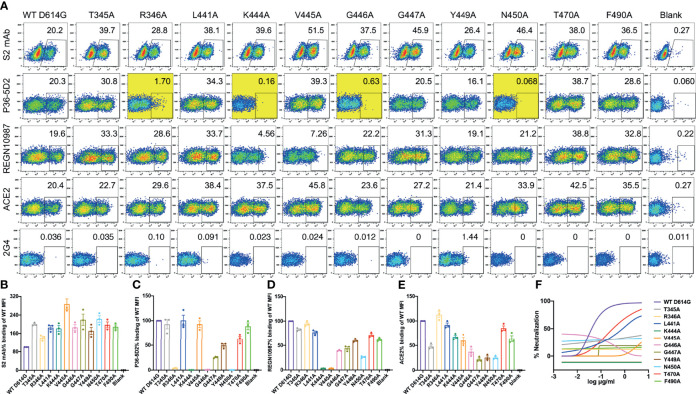
As severe acute respiratory syndrome coronavirus 2 (SARS-CoV-2) variants continue to emerge and spread around the world, antibodies and vaccines to confer broad and potent neutralizing activity are urgently needed. Through the isolation and characterization of monoclonal antibodies (mAbs) from individuals infected with SARS-CoV-2, we identified one antibody, P36-5D2, capable of neutralizing the major SARS-CoV-2 variants of concern. Crystal and electron cryo-microscopy (cryo-EM) structure analyses revealed that P36-5D2 targeted to a conserved epitope on the receptor-binding domain of the spike protein, withstanding the three key mutations-K417N, E484K, and N501Y-found in the variants that are responsible for escape from many potent neutralizing mAbs, including some already approved for emergency use authorization (EUA). A single intraperitoneal (IP) injection of P36-5D2 as a prophylactic treatment completely protected animals from challenge of infectious SARS-CoV-2 Alpha and Beta. Treated animals manifested normal body weight and were devoid of infection-associated death up to 14 days. A substantial decrease of the infectious virus in the lungs and brain, as well as reduced lung pathology, was found in these animals compared to the controls. Thus, P36-5D2 represents a new and desirable human antibody against the current and emerging SARS-CoV-2 variants.

Keywords: SARS-CoV-2; epitope; human neutralizing antibody; in vivo protection; variants of concern.


Conflict of interest statement

The authors declare that the research was conducted in the absence of any commercial or financial relationships that could be construed as a potential conflict of interest.

Figures

Figure 1
Figure 1
P36-5D2 shows broad and potent neutralizing and binding spectrum against pseudotyped and infectious SARS-CoV-2. P36-5D2, P36-1B7, P74-6D2, and P36-1A3 were the top 4 isolated neutralizing monoclonal antibodies (mAbs) against severe acute respiratory syndrome coronavirus 2 (SARS-CoV-2) from infected individuals. Published representative receptor-binding domain (RBD)-specific mAbs included REGN10933 (class I), CB6 (class I), BD356-2 (class II), and REGN10987 (class III). The neutralizing and binding ability of mAbs against pseudotyped SARS-CoV-2, including Alpha (B.1.1.7), Beta (B.1.351), and Gamma (P.1), and the respective mutations derived from the three variants of concern (VOCs) were evaluated. Values indicate the fold changes in half-maximal inhibitory concentrations (IC50) (A) and the mean fluorescence intensity (MFI) relative to that of wild-type (WT) D614G (B). The IC50 of antibodies against WT D614G are listed in (A). The minus symbol represents increased resistance and the plus sign indicates increased sensitivity. IC50 or MFI highlighted in red indicates that resistance increased at least threefold; in blue, sensitivity increased at least threefold. BDL (below detection limit) represents the highest concentration of mAbs that failed to reach 50% potency in neutralization activity or mAbs that failed to bind the cell surface-expressed SARS-CoV-2 variants. Results are presented as the mean value from three independent experiments. (C) Neutralization of P36-5D2 against the infectious SARS-CoV-2 WT, Alpha, and Beta variants. 2G4 is the negative control antibody specific for Ebola virus glycoprotein (EBOV GP). Experiments were performed in triplicate, and all data were presented as the means ± SEM.
Figure 2
Figure 2
P36-5D2 binds to a highly conserved epitope on the receptor-binding domain (RBD) and avoids three key mutant residues: K417N, E484K, and N501Y. (A) The epitope of P36-5D2 (purple) is highlighted on the surface of severe acute respiratory syndrome coronavirus 2 (SARS-CoV-2) RBD-3M carrying K417T, E484K, and N501Y (cyan) (PDB: 7FJC). RBD-3M residues on the epitope are labeled. The percentages show the conservation at these positions observed in the GISAID EpiCoV database by aligning 1,959,333 sequences of the SARS-CoV-2 spike from December 2019 to July 2021. (B) The paratope of P36-5D2 (sliver) is highlighted on heavy chain (purple) and light chain (pink). Antibody residues are labeled in purple or pink depending on their origin from heavy or light chain. (C, D) Interaction between P36-5D2 Fab and SARS-CoV-2 RBD-3M. (E–H) Listed class III mAbs Fab fragments are superposed onto P36-5D2/RBD-3M crystal structure. REGN10987, S309, C110, and C135 are reference class III mAbs. The epitopes of these antibodies were assigned by selection of any RBD residue within 4 Å of any antibody residue. The epitope of REGN10987 (PDB: 6XDG) is colored in blue, S309 (PDB: 7JX3) in red, C110 (PDB: 7K8V) yellow, C135 (PDB: 7K8Z) green, and P36-5D2 in purple. (I) Electron cryo-microscopy (cryo-EM) for the P36-5D2 Fab and SARS-CoV-2 spike complex (left, 3.65 Å; right, 3.69 Å) reveals the binding of P36-5D2 Fab to both “up” and “down” RBDs. P36-5D2/RBD-3M crystal structure shown as cartoon superposed onto one protomer of P36-5D2/spike cryo-EM structure. The spike is shown as a molecular surface, with one protomer RBD colored cyan, NTD green, SD1-2 yellow, and S2 gray. The top view shows that P36-5D2 avoids residues K417, E484, and N501, which are shown as red-colored spheres. Inset: structure superposition showing clashes between ACE2 (PDB: 6MOJ; green) and P36-5D2 Fab. Overlapping residues shared between P36-5D2 and ACE2 are listed below.
Figure 3
Figure 3
Impact of single alanine mutated residues on P36-5D2 binding and neutralization. (A) Wild-type and single alanine mutated spike were expressed on the surface of HEK 293T cells, incubated with P36-5D2, REGN10987, or ACE2, followed by staining with anti-human IgG Fc phycoerythrin (PE) or anti-His PE, and analyzed by FACS. For each panel, the X-axis means tested antibody or ACE2 binding PE/FITC and the Y-axis means side-scattered light (SSC). The gated cell percentages are shown. (B) The S2 monoclonal antibody (mAb) is a positive control antibody used for spike expression normalization. (C–E) The relative mean fluorescence intensity (MFI) of mAbs or ACE2 binding was determined by comparing the total MFI in the selected gate between the spike variants and WT D614G. 2G4 targeting Ebola virus glycoprotein (EBOV GP) is a negative control mAb. NC denotes HEK 293T cells with mock transfection. Alanine mutated residues completely destroying P36-5D2 binding are highlighted in yellow. Data are presented as the mean ± SEM from three independent experiments. (F) Impact of single alanine mutated residues on pseudovirus neutralization sensitivity to P36-5D2. Results are presented as the mean value from three independent experiments.
Figure 4
Figure 4
Efficacy of P36-5D2 prophylaxis against the infectious severe acute respiratory syndrome coronavirus 2 (SARS-CoV-2) Alpha or Beta variant in K18-hACE-2 mice. (A) Experimental schedule for antibody prophylaxis. Eight-week-old K18-hACE2 transgenic female mice were administered 10 mg/kg of P36-5D2 intraperitoneally or untreated 1 day prior to challenge with 103 plaque-forming units (PFU) infectious SARS-CoV-2 Alpha or Beta via the intranasal route. (B–E) The survival percentage (B, D) and body weight (C, E) were recorded daily after infection until the occurrence of death or until the experimental end point at 14 days post-infection (dpi) (P36-5D2, n = 6; untreated, n = 5). Mice were sacrificed at 4 dpi for virus titer analysis (P36-5D2, n = 6; untreated, n = 7). (F–I) Lung titers (F, H) and brain titers (G, I) were tested by plaque assays in lung and brain tissue homogenates. The PFU per tissue were compared between groups in log10-transformed units. All data are presented as the mean ± SEM. Analysis of a two-tailed unpaired t-test was used. *p < 0.05, **p < 0.01. n.s., not significant. (J, K) H&E staining of lung sections from P36-5D2-injected or untreated mice at 4 dpi. VL, vascular lumen; BL, bronchiolar lumen. Scale bars, 50 mm. Each image is representative of each group.

Similar articles

Cited by

References

    1. Fujino T, Nomoto H, Kutsuna S, Ujiie M, Suzuki T, Sato R, et al. . Novel SARS-CoV-2 Variant in Travelers From Brazil to Japan. Emerg Infect Dis (2021) 27(4):1243–5. doi: 10.3201/eid2704.210138 - DOI - PMC - PubMed
    1. Kupferschmidt K. Fast-Spreading U.K. Virus Variant Raises Alarms. Science (2021) 371(6524):9–10. doi: 10.1126/science.371.6524.9 - DOI - PubMed
    1. Maggi F, Novazzi F, Genoni A, Baj A, Spezia PG, Focosi D, et al. . Imported SARS-CoV-2 Variant P.1 in Traveler Returning From Brazil to Italy. Emerg Infect Dis (2021) 27(4):1249–51. doi: 10.3201/eid2704.210183 - DOI - PMC - PubMed
    1. Tegally H, Wilkinson E, Giovanetti M, Iranzadeh A, Fonseca V, Giandhari J, et al. . Detection of a SARS-CoV-2 Variant of Concern in South Africa. Nature (2021) 592(7854):438–43. doi: 10.1038/s41586-021-03402-9 - DOI - PubMed
    1. Tegally H, Wilkinson E, Lessells RJ, Giandhari J, Pillay S, Msomi N, et al. . Sixteen Novel Lineages of SARS-CoV-2 in South Africa. Nat Med (2021) 27(3):440–6. doi: 10.1038/s41591-021-01255-3 - DOI - PubMed

Publication types

Supplementary concepts