. 2022 Apr;31(4):811-821.
doi: 10.1002/pro.4269. Epub 2022 Jan 12.

Completing the family of human Eps15 homology domains: Solution structure of the internal Eps15 homology domain of γ-synergin

Affiliations

Completing the family of human Eps15 homology domains: Solution structure of the internal Eps15 homology domain of γ-synergin

Michael Kovermann et al. Protein Sci. 2022 Apr.

Abstract

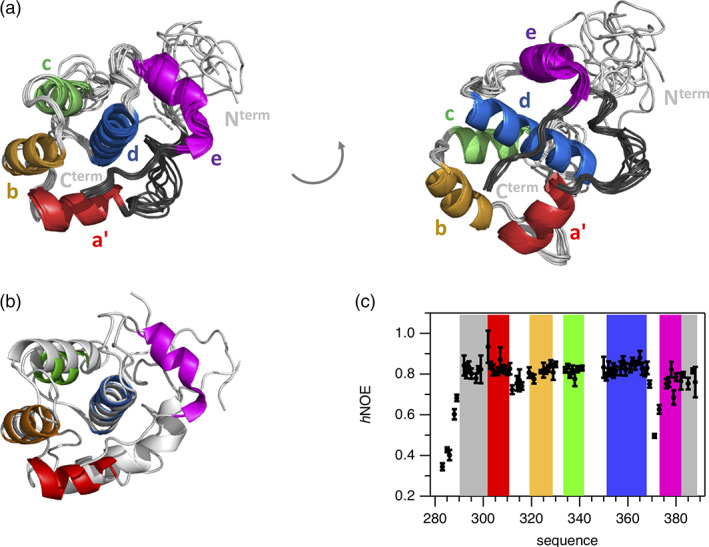
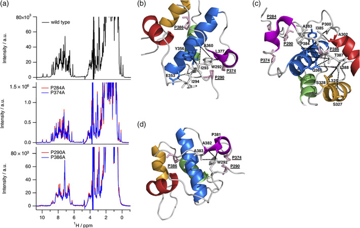
Eps15 homology (EH) domains are universal interaction domains to establish networks of protein-protein interactions in the cell. These networks mainly coordinate cellular functions including endocytosis, actin remodeling, and other intracellular signaling pathways. They are well characterized in structural terms, except for the internal EH domain from human γ-synergin (EHγ). Here, we complete the family of EH domain structures by determining the solution structure of the EHγ domain. The structural ensemble follows the canonical EH domain fold and the identified binding site is similar to other known EH domains. But EHγ differs significantly in the N- and C-terminal regions. The N-terminal α-helix is shortened compared to known homologues, while the C-terminal one is fully formed. A significant proportion of the remaining N- and C-terminal regions are well structured, a feature not seen in other EH domains. Single mutations in both the N-terminal and the C-terminal structured extensions lead to the loss of the distinct three-dimensional fold and turn EHγ into a molten globule like state. Therefore, we propose that the structural extensions in EHγ function as a clamp and are undoubtedly required to maintain its tertiary fold.

Keywords: EF hand; EH domain; Eps15 homology domain; NMR spectroscopy; molten globule; structure determination.


Conflict of interest statement

Authors declare that they have no competing interests.

Figures

FIGURE 1
FIGURE 1
Composition of the EH𝛾 domain and initial screen of potential interaction to different SCAMP1 constructs. (a) Primary outline of the EH𝛾 domain comprising structural elements a′, b, c, d, and e determined as α‐helical in this study as well as structured N‐ and C‐terminal extensions that are highlighted in dark‐gray (see also Figure 2). The N‐terminal SCAMP1 construct is devoid of any transmembrane helices and comprises two domains that can be divided into a coiled coil (SCAMP1′) and NPF repeat rich motifs (SCAMP1″). SCAMP1″ has been further subdivided into peptide A, B and C in this study enabling to probe individual interactions to the EH𝛾 domain. The primary sequences of all molecules probed in this study are shown in SI Table 1. (b) Probing the interaction between the EH𝛾 domain and SCAMP1″ applying analytical size exclusion chromatography. The formed complex (continuous line, colored in red) eluates at a lower retention volume compared to the isolated EH𝛾 domain (dashed line, colored in blue) and SCAMP″ (dotted line, colored in orange). (c) Following the interaction between the EH𝛾 domain and SCAMP1″ using isothermal titration calorimetry (ITC). The results of the quantitative analyses applying a single site binding model are presented right to the ITC profile, n.b. refers to no binding
FIGURE 2
FIGURE 2
Structural and dynamical characterization of the EH𝛾 domain. (a) The three‐dimensional structure of the EH𝛾 domain is shown as an alignment of the 10 lowest energy structures. α‐Helices are color‐coded and labeled. Also, well‐structured regions N‐terminal of α‐helix a′ and C‐terminal of helix e have been colored (dark‐gray). Accompanying SI Table 3 lists NMR restraints and statistical analysis of the NMR‐based structure calculation. (b) Comparison between the lowest energy structure of EH𝛾 determined in this study (helical elements highlighted in color) to homologues EH domain of EHD1 possessing pdb code 2JQ6 (colored in light grey). The alignment has been conducted for backbone atoms comprising helix d (K352‐R367 in EH𝛾 and D103‐E118 in EHD1). Further individual comparisons between EH𝛾 and homologues structures are shown in SI Figure 4. (c) {1H‐}15N heteronuclear NOE ( h NOE) acquired for the EH𝛾 domain at T = 298 K and B 0 = 20 T reporting on backbone dynamics on the pico‐to‐nanosecond time scale. Colors used for highlighting the background refer to the structural composition presented in panel a. Error bars refer to the standard deviation obtained from three independent measurements
FIGURE 3
FIGURE 3
Interaction between the EH𝛾 domain and peptides comprising the NPF motif found in SCAMP1. (a) Analysis of changes of chemical shifts of NMR resonance signals following the interaction between isotopically labeled EH𝛾 and peptide A (top), B (mid), and C (bottom). Colors used for highlighting the background refer to the structural composition of EH𝛾 presented in Figure 2a. Values for Δ𝜔 have been calculated using the molar ratio n = 2 between peptide and EH𝛾, respectively. The cutoff value identifying most affected residues has been set for all titration experiments to Δ𝜔 = 0.055 ppm. (b) Overlay of two‐dimensional heteronuclear 1H‐15N HSQC NMR spectra following the interaction between peptide A and EH𝛾 acquired for different stoichiometric ratios at T = 298 K and B 0 = 20 T: n = 0 (colored in red), n = 0.4 (colored in orange), n = 0.8 (colored in magenta), n = 2 (colored in blue). Residues of EH𝛾 that are most affected upon binding are labeled by using the one letter code for amino acids followed by the position in the primary sequence, sc refers to side chain. (c) Highlighting all residues of EH𝛾 exceeding Δ𝜔 = 0.055 ppm in all three titration experiments shown in panel A. These residues are presented including side chains (stick mode) in pink and labeled. (d) Individual titration profiles observed for 1H‐15N correlations of I324 (colored in red), L325 (colored in blue), and W339 (colored in orange) of EH𝛾 when peptide A (closed symbols), peptide B (open symbols), or peptide C (symbols with inner dot) has been stepwise added. The binding affinity has been determined to K D pepA = 110 ± 10 μM (continuous line), K D pepB = 120 ± 30 μM (dashed line), K D pepC = 30 ± 20 μM (dot‐dash line) by applying a joint fitting procedure for these residues to an one site binding model
FIGURE 4
FIGURE 4
Conformational heterogeneity of EH𝛾. (a) Section of two‐dimensional heteronuclear 1H‐15N HSQC NMR spectrum acquired for EH𝛾 reveals at least two conformations that possess different chemical shifts for a distinct set of residues (cross‐peaks arising from the minor conformation are indicated by “b” following the position in the primary sequence). (b–e) Characterization of two conformations observed for EH𝛾 and two variants P284A and P374A regarding differences in chemical shifts (b) and in the ratio of signal heights (c–e) observed in an 1H‐15N‐HSQC NMR spectrum. Colors used for highlighting the background refer to the structural composition of EH𝛾 presented in Figure 2a. Please note that no spectroscopic information could be obtained for A285 and Q286 (P284A variant) as for A376 (P374A variant)
FIGURE 5
FIGURE 5
Structural impact of selected proline residues comprising EH𝛾. (a) Overlay of one‐dimensional proton NMR spectra acquired for wild type (top), P284A (colored in red) and P374A (colored in blue) (mid) as well as P290A (colored in red) and P386A (colored in blue) protein variants (bottom). (b) Network of NOEs illustrating structural contacts between residues comprising helices d (colored in blue), e (colored in magenta) and N‐terminal residues. (c) Network of NOEs illustrating structural contacts between residues comprising helices a’ (colored in red), b (colored in orange), d (colored in blue) and C‐terminal residues. (d) Network of NOEs illustrating structural contacts between N‐ and C‐terminal residues. Proline residues used for particular alanine replacement are shown with side chains (stick mode, colored in pink) and have been underlined. NOE contacts have been highlighted by dotted lines (colored in black)

Similar articles

References

    1. Fazioli F, Minichiello L, Matoskova B, Wong WT, Di Fiore PP. eps15, a novel tyrosine kinase substrate, exhibits transforming activity. Mol Cell Biol. 1993;13(9):5814–5828. - PMC - PubMed
    1. Wong WT, Kraus MH, Carlomagno F, et al. The human eps15 gene, encoding a tyrosine kinase substrate, is conserved in evolution and maps to 1p31‐p32. Oncogene. 1994;9(6):1591–1597. - PubMed
    1. Wong WT, Schumacher C, Salcini AE, et al. A protein‐binding domain, EH, identified in the receptor tyrosine kinase substrate Eps15 and conserved in evolution. Proc Natl Acad Sci U S A. 1995;92(21):9530–9534. - PMC - PubMed
    1. Confalonieri S, Di Fiore PP. The Eps15 homology (EH) domain. FEBS Lett. 2002;513(1):24–29. - PubMed
    1. Salcini AE, Confalonieri S, Doria M, et al. Binding specificity and in vivo targets of the EH domain, a novel protein‐protein interaction module. Genes Dev. 1997;11(17):2239–2249. - PMC - PubMed

Publication types

MeSH terms

Substances