. 2022 Jan 25;50(2):899-914.
doi: 10.1093/nar/gkab1253.

Redirecting meiotic DNA break hotspot determinant proteins alters localized spatial control of DNA break formation and repair

Affiliations

Redirecting meiotic DNA break hotspot determinant proteins alters localized spatial control of DNA break formation and repair

Randy W Hyppa et al. Nucleic Acids Res. .

Abstract

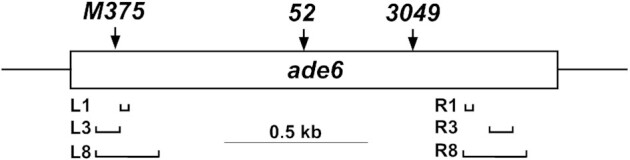
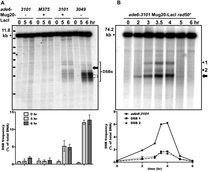
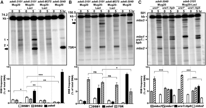
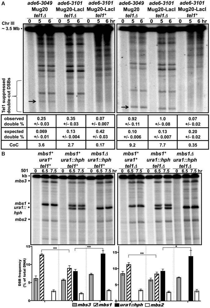
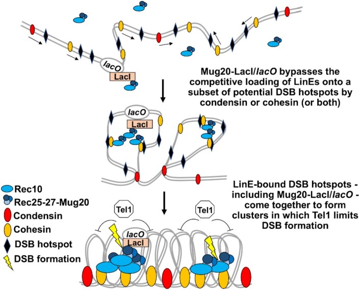
During meiosis, DNA double-strand breaks (DSBs) are formed at high frequency at special chromosomal sites, called DSB hotspots, to generate crossovers that aid proper chromosome segregation. Multiple chromosomal features affect hotspot formation. In the fission yeast S. pombe the linear element proteins Rec25, Rec27 and Mug20 are hotspot determinants - they bind hotspots with high specificity and are necessary for nearly all DSBs at hotspots. To assess whether they are also sufficient for hotspot determination, we localized each linear element protein to a novel chromosomal site (ade6 with lacO substitutions) by fusion to the Escherichia coli LacI repressor. The Mug20-LacI plus lacO combination, but not the two separate lac elements, produced a strong ade6 DSB hotspot, comparable to strong endogenous DSB hotspots. This hotspot had unexpectedly low ade6 recombinant frequency and negligible DSB hotspot competition, although like endogenous hotspots it manifested DSB interference. We infer that linear element proteins must be properly placed by endogenous functions to impose hotspot competition and proper partner choice for DSB repair. Our results support and expand our previously proposed DSB hotspot-clustering model for local control of meiotic recombination.


Figures

Figure 1.
Figure 1.
Location of ade6 mutations. lacO operator arrays (horizontal brackets) of 1, 3 or 8 copies were substituted for an identical size interval of the ade6 open reading frame (open box; 1659 bp) on the left (L) at the ade6-M375 position (single bp control mutation G133T, three bp from the ade6-M26 recombination hotspot with ATGACGT bound by the transcription factor Atf1-Pcr1), or on the right (R). The ade6-52 single bp mutation (G796A), between the L and R arrays, was crossed with the arrays to determine intragenic recombinant frequencies. ade6-3049 (C1214T) creates another ATGACGT heptamer and a strong DSB hotspot. arg1, 300 kb to the right, was used to determine intergenic recombinant frequencies. bub1-243, 0.65 kb to the left of ade6, and vtc4-1104, 1.49 kb to the right, were used for DNA analyses of recombinants and intermediates (Figure 6).
Figure 2.
Figure 2.
Linear element fusion protein Mug20-LacI induces abundant DSBs at the ade6-3101 hotspot. (A) Formation and accumulation of DSBs in rad50S strains with ade6-3101 containing 8 lacO operators (at the thick black arrow) alone, with Mug20-LacI alone (mug20-231), or with both to generate the ade6-3101 hotspot. ade6-3049 (thick white arrow) is a non-LacI-lacO DSB hotspot control. Bracket indicates the ∼2 kb region of DSB formation in the two strains. Cells were induced for meiosis and harvested at the indicated times. DNA was digested with BsrGI and analyzed by electrophoresis and Southern blot hybridization using a probe at the right end of the 11.8 kb fragment with ade6. Black ovals on the left margin indicate a DNA ladder (1 kb Plus, Invitrogen; from the top 15, 10, 8, 7, 6, 5, 4, and 3 kb). Quantification is based on 2 or 3 blots from two independent inductions; error bars indicate the range or SEM. (B) Early formation and timely repair of DSBs in a rad50+ strain with ade6-3101 and Mug20-LacI (mug20-231). DNA was analyzed as in (A) after digestion with PmeI using a probe at the right end of the 74.2 kb fragment with ade6. DSBs at ade6-3101 are indicated by the thick black arrow (∼20 kb fragment); endogenous DSB sites 1 and 2 are 15 and 5 kb from ade6. Quantification is based on two independent inductions; error bars (some invisible) indicate the range. Note that DSBs at ade6-3101 are visible before replication is complete at 3 h, but DSBs at endogenous site 1, site 2, and mbs1 are not (see also Supplementary Figure S1).
Figure 3.
Figure 3.
The Mug20-LacI fusion protein lacks DSB hotspot competition at the ade6-3101 hotspot but retains competition at the endogenous mbs1 hotspot. rad50S strains were induced for meiosis, and DNA analyzed as in Figure 2. In each panel, quantification is based on n blots from two independent inductions; error bars indicate SEM (range for n = 2). (A) ade6-3049 but not ade6-3101 plus Mug20-LacI (mug20-231) competes with close hotspots; ade6-M375 is a non-hotspot control. DNA was digested with PmeI and analyzed on three to five blots with a probe at the right end of the 74.2 kb fragment containing ade6. DSBs at sites 1 and 2 were significantly less frequent with ade6-3049 than with ade6-M375 (***P < 0.0001 for site 1 and **P = 0.0004 for site 2) but were not less frequent with the ade6-3101 hotspot (thick black arrow, ∼20 kb). Rather, DSBs at site 1 were moderately stimulated by ade6-3101 plus Mug20-LacI (mug20-231) versus ade6-3101 alone or M375: *P = 0.011 or **0.004, respectively). DSBs at site 2 were not significantly different (P = 0.18 or 0.054, respectively). (B) ade6-3049 but not the ade6-3101 hotspot (DSBs indicated by thick black arrow, ∼110 kb) competes with a distant hotspot. DNA was digested with SacII and analyzed on three or four blots with a probe at the right end of the 150.5 kb fragment with ade6. Only ade6-3049 competed with the strong DSB hotspot 75 kb to the right of ade6 (75R) (*P = 0.015) or with weaker DSBs in between. (C) Mug20-LacI manifests DSB competition on another chromosome. DNA was digested with NotI and analyzed on four to seven blots with a probe at the left end of the 501 kb NotI fragment J. mbs1 was competed by the artificial hotspot ura1::hph in both Mug20 and Mug20-LacI strains (***P < 0.0001). mbs2, an endogenous hotspot 100 kb to the left of mbs1, was also competed by ura1::hph in Mug20-LacI strains (***P < 0.0001). DSBs at mbs3, 200 kb to the right of mbs1, did not differ significantly in these strains. Strains with no black bar (first and third from the left) are ura1+.
Figure 4.
Figure 4.
The ade6-3101 hotspot manifests Tel1-dependent DSB interference; Tel1-independent DSB competition indicates separate mechanisms. rad50S strains were induced for meiosis, and DNA analyzed as in Figure 3, except meiosis was at 25°C in (B). (A) Both ade6-3049 and ade6-3101 manifest Tel1-dependent DSB interference. DNA was digested with NotI and analyzed on four to six blots with a probe between ade6 and the 75R DSB hotspot (left panel) or between ade6 and the tel1L hotspot near tel1 (right panel). Double-cut DSBs (black arrows; 75 kb left and 40 kb right) were evident in tel1Δ (left and middle lane sets) but not in tel1+ (right lane set). Coefficients of coincidence (CoC; mean ± SEM) show positive DSB interference (1 - CoC) in tel1+ and negative interference in tel1Δ. Single-cut DSBs and frequencies are visible in Supplementary Figure S3 using a different radioactive probe. (B) DSB competition at mbs1 is Tel1-independent. DNA was digested with NotI and analyzed on three to seven blots with a probe at the left end of the 501 kb NotI fragment J. DSBs at both mbs1+ and ura1::hph hotspots were reduced in the presence of the other hotspot (compare the double hotspot in the middle lane set to either single hotspot; **P = 0.007 for mbs1+ and **P = 0.0034 for ura1::hph), indicating mutual DSB competition. This competition was also present without Tel1 (right panel; **P = 0.002 for mbs1+ and *P = 0.018 for ura1::hph).
Figure 5.
Figure 5.
Joint DNA molecules arise at similar time and frequency at the ade6-3101 and ade6-3049 DSB hotspots. mus81Δ strains were induced for meiosis, and DNA, digested with BsrGI, was analyzed by two-dimensional gel electrophoresis and Southern blot hybridization using a probe near the right end of the 11.8 kb fragment with ade6. (A) Branched DNA molecules, predominantly replication intermediates (Y-shaped; thick white arrows), arose at 2 h, and recombination intermediates (X-shaped Holliday junctions; thin black arrows) appeared at 4–6 h. The prominent spot is the parental DNA fragment. (B) Expanded view of replication arc at 2 h, showing a prominent pause or DSB site in the ade6-3101 strain (bottom panel) but not in the ade6-3049 strain (upper panel). (C) For quantification, branched DNA (structures above the linear DNA arc) was normalized to total DNA. Quantification is based on two or three blots from two independent inductions; error bars indicate the range or SEM.
Figure 6.
Figure 6.
Less interhomolog DSB repair occurs at the ade6-3101 hotspot than at the ade6-3049 hotspot, and crossover DNA is strongly reduced.Diploid strains heterozygous for bub1-243 (L) and vtc4-1104 (R), flanking ade6 were used to distinguish intersister (IS) and interhomolog (IH) Holliday junctions (HJs) in (A) and crossover DNA in (B) (53). (A) mus81Δ strains were induced for meiosis and harvested at 5 h. DNA was digested with PmlI and ScaI and analyzed as in Figure 5 using a probe near the middle of ade6. IS HJs (black arrows) and IH HJs (white arrows) were quantified from three (ade6-3049) or six (ade6-3101 mug20-231) blots from two independent inductions; data are IS L/P1, IS R/P2, and IH/[(P1 + P2)/2], each as % ± SEM, where P1 and P2 are parental DNAs 1 and 2, respectively. The IS to IH ratio of HJs is indicated. (B) Strains with the indicated homozygous ade6 alleles were induced for meiosis and harvested at 5 hr; DNA was analyzed as in (A), except electrophoresis was in only one dimension. ade6-3057 is a non-hotspot control (Figure 1). R1 and R2 are reciprocal recombinant fragments. The fraction of crossover fragment, 2 × R2/(P1 + P2) because R1 can also arise from a partial restriction digestion, is based on three to five blots from two independent meiotic inductions; error bars indicate the SEM.
Figure 7.
Figure 7.
Model for DSB competition arising from the competitive loading of LinE complexes onto DSB hotspots. A loader, such as cohesin or condensin, moves along paired sister chromatids (thin black arrows) and loads LinE complexes (blue circles and ovals) onto a limited number of potential DSB hotspot sites. This prevents other sites in this traversed interval from being bound by LinEs and therefore limits DSBs to only the LinE-loaded sites (DSB competition). The ade6-3101 hotspot with a lacO array allows independent loading of Mug20-LacI and thus lacks DSB competition. The loader (cohesin or condensin) groups the LinE-hotspot complexes, including the Mug20-LacI-bound site, into a cluster, in which a DSB is formed. This DSB activates Tel1 protein kinase to prevent further DSB formation in that cluster (DSB interference).

References

    1. Zickler D., Kleckner N.. Meiotic chromosomes: integrating structure and function. Annu. Rev. Genet. 1999; 33:603–754. - PubMed
    1. Bhalla N., Dernburg A.F.. Prelude to a division. Annu. Rev. Cell Dev. Biol. 2008; 24:397–424. - PMC - PubMed
    1. Nicklas R.B., Koch C.A.. Chromosome micromanipulation. 3. Spindle fiber tension and the reorientation of mal-oriented chromosomes. J. Cell Biol. 1969; 43:40–50. - PMC - PubMed
    1. Lam I., Keeney S.. Mechanism and regulation of meiotic recombination initiation. Cold Spring Harb. Perspect. Biol. 2015; 7:a016634. - PMC - PubMed
    1. Nambiar M., Chuang Y.C., Smith G.R.. Distributing meiotic crossovers for optimal fertility and evolution. DNA Repair (Amst.). 2019; 81:102648. - PMC - PubMed

Publication types

MeSH terms

Substances