Review
. 2022 Jan 25;50(2):617-634.
doi: 10.1093/nar/gkab1256.

Reexamining assumptions about miRNA-guided gene silencing

Affiliations
Review

Reexamining assumptions about miRNA-guided gene silencing

Audrius Kilikevicius et al. Nucleic Acids Res. .

Abstract

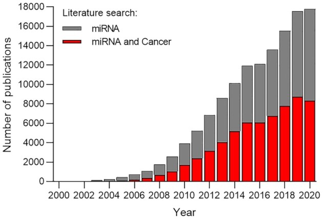
MicroRNAs (miRNAs) are short endogenously expressed RNAs that have the potential to regulate the expression of any RNA. This potential has led to the publication of several thousand papers each year connecting miRNAs to many different genes and human diseases. By contrast, relatively few papers appear that investigate the molecular mechanism used by miRNAs. There is a disconnect between rigorous understanding of mechanism and the extraordinary diversity of reported roles for miRNAs. Consequences of this disconnect include confusion about the assumptions underlying the basic science of human miRNAs and slow development of therapeutics that target miRNAs. Here, we present an overview of investigations into miRNAs and their impact on gene expression. Progress in our understanding of miRNAs would be aided by a greater focus on the mechanism of miRNAs and a higher burden of evidence on researchers who seek to link expression of a particular miRNA to a biological phenotype.


Figures

Figure 1.
Figure 1.
Biogenesis of miRNAs. (A) A miRNA is transcribed into pri-miRNA by Pol II. (B) Drosha/DCGR8 microprocessor complex cleaves the pri-miRNA to pre-miRNA, which enters the cytoplasm. (C) The mature duplex miRNA is generated by Dicer.
Figure 2.
Figure 2.
Interactions between AGO protein, an miRNA guide strand, and an RNA target. AGO proteins are composed of four domains: The N-terminal domain supports miRNA loading, the PAZ domain anchors the 3′ end while the MID domain binds the 5′ end of the miRNA. (A) Argonaute loaded miRNA seed region and other regions. (B) PIWI is the protein domain responsible for cleaving RNA substrates by AGO2 when there is full complementarity. (C) Centrally mismatched bases will block cleavage while permitting binding of a miRNA to a target RNA. PAZ – PIWI-Argonaute-Zwille, N – N-terminal (amino-terminal), PIWI – P-element-induced whimpy testes and MID – middle domains.
Figure 3.
Figure 3.
miRNA families. (A) Six miRNA families account for ∼50% of top 100 miRNAs loaded on AGO2 in HCT116 cells (60). (B) miR-96 is the only member of its family while Let-7 has several family members expressed. (C) Sequence variation among Let-7–5p/98–5p family members.
Figure 4.
Figure 4.
An example of the impact of cooperative binding on repression of gene expression by duplex RNAs. In this example an anti-CAG repeat duplex RNA is introduced into patient-derived Huntington's disease cells. (A) The wild-type huntingtin (HTT) allele has 17 repeats, while the mutant allele has 69 repeats. 1–2 anti-CAG small RNAs can bind to the wild-type allele while as many as nine anti-CAG RNAs can bind to the mutant allele. (B) Allele selective inhibition by a small RNA with a central mismatch relative to the RNA target.
Figure 5.
Figure 5.
Single versus multiple binding of miRNAs and cooperativity. (A) Binding of a single miRNA to a target, no potential for cooperativity. Multiple miRNAs of the (B) same or (C) different miRNA families. (D) Bridging of AGO:miRNA complexes by TNRC6 scaffolding protein provides a structural basis for cooperativity.
Figure 6.
Figure 6.
Interplay of miRNA and target concentration. miR-122 was prioritized for study because of (A) high expression in hepatocyte cells relative to most other miRNAs and (B) other cell types. (C) Model showing the correlation between miRNA concentration, target mRNA concentration, and potential for biologically significant inhibition of gene expression. CPM – counts per million. Data obtained from FANTOM5 database (77).
Figure 7.
Figure 7.
Anti-miRs, tools for investigating the function of miRNAs. When an anti-miR is present it has the potential to block the miRNA and prevent miRNA-mediated repression of the target RNA.
Figure 8.
Figure 8.
Challenges to identifying candidate miRNAs from CLIP data and predictions programs. (A, B) Show AGO2 eCLIP data (60) in HCT116 cells for the MYC 3′-UTR. miRNAs were predicted using (A) TargetScan or (B) miRANDA. (C) Let-7f, can example of a highly expressed miRNA that was not predicted by TargetScan but was predicted by miRANDA. Red labeled miRNAs significantly loaded on AGO2 over AGO2 KO cell line.
Figure 9.
Figure 9.
The miRNA literature. Publications identified by PubMed searches for the terms ‘miRNA’ and ’miRNA and cancer’, 2000–2020.

References

    1. Bartel D.P. Metazoan microRNAs. Cell. 2018; 173:20–51. - PMC - PubMed
    1. Treiber T., Treiber N., Meister G.. Regulation of microRNA biogenesis and its crosstalk with other cellular pathways. Nat. Rev. Mol. Cell Biol. 2019; 20:5–20. - PubMed
    1. Banerjee D., Slack F.. Control of developmental timing by small temporal RNAs: a paradigm for RNA-mediated regulation of gene expression. Bioessays. 2002; 24:119–129. - PubMed
    1. Pasquinelli A.E., Ruvkun G.. Control of developmental timing by micrornas and their targets. Annu. Rev. Cell Dev. Biol. 2002; 18:495–513. - PubMed
    1. Lee R.C., Feinbaum R.L., Ambros V. The C. elegans heterochronic gene lin-4 encodes small RNAs with antisense complementarity to lin-14. Cell. 1993; 75:843–854. - PubMed

Publication types