

Dynamic functional connectivity in modular organization of the hippocampal network marks memory phenotypes in temporal lobe epilepsy
- PMID: 34967488
- PMCID: PMC8933317
- DOI: 10.1002/hbm.25763
Dynamic functional connectivity in modular organization of the hippocampal network marks memory phenotypes in temporal lobe epilepsy
Abstract
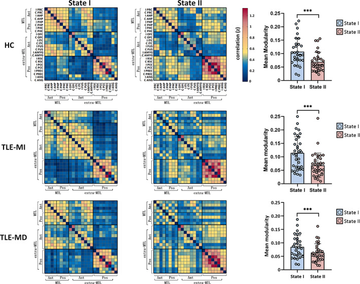
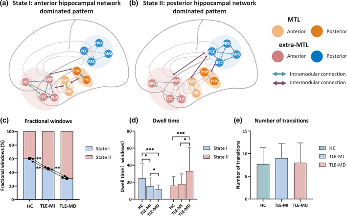
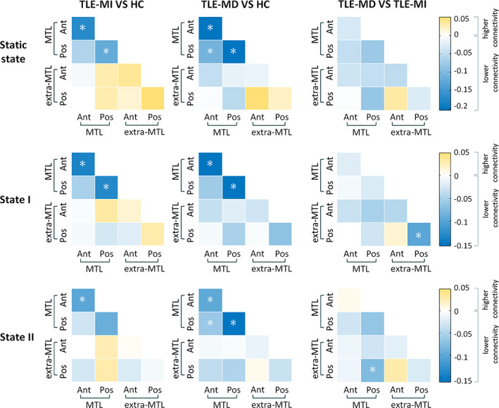
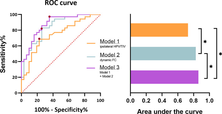
Temporal lobe epilepsy (TLE) is a network disorder with a high incidence of memory impairment. Memory processing ability highly depends on the dynamic coordination between distinct modules within the hippocampal network. Here, we investigate the relationship between memory phenotypes and modular alterations of dynamic functional connectivity (FC) in the hippocampal network in TLE patients. Then, 31 healthy controls and 66 TLE patients with hippocampal sclerosis were recruited. The patients were classified into memory-intact (MI, 35 cases) group and memory-deficit (MD, 31 cases) group, each based on individual's Wechsler Memory Scale-Revised score. The sliding-windows approach and graph theory analysis were used to analyze the hippocampal network based on resting state functional magnetic resonance imaging. Temporal properties and modular metrics were calculated. Two discrete and switchable states were revealed: a high modularized state (State I) and a low modularized state (State II), which corresponded to either anterior or posterior hippocampal network dominated pattern. TLE was prone to drive less State I but more State II, and the tendency was more obvious in TLE-MD. Additionally, TLE-MD showed more widespread alterations of modular properties compared with TLE-MI across two states. Furthermore, the dynamic modularity features had unique superiority in discriminating TLE-MD from TLE-MI. These findings demonstrated that state transitions and modular function of dissociable hippocampal networks were altered in TLE and more importantly, they could reflect different memory phenotypes. The trend revealed potential values of dynamic FC in elucidating the mechanism underlying memory impairments in TLE.
Keywords: MRI; chronic epilepsy; dynamics; hippocampal sclerosis; memory disorder; neuroimaging.
© 2021 The Authors. Human Brain Mapping published by Wiley Periodicals LLC.
Conflict of interest statement
The authors declare no conflicts of interest.
Figures
Similar articles
-
Comparision of spontaneous brain activity between hippocampal sclerosis and MRI-negative temporal lobe epilepsy.Epilepsy Behav. 2024 Aug;157:109751. doi: 10.1016/j.yebeh.2024.109751. Epub 2024 May 30. Epilepsy Behav. 2024. PMID: 38820678
-
Differential sensitivity of structural, diffusion, and resting-state functional MRI for detecting brain alterations and verbal memory impairment in temporal lobe epilepsy.Epilepsia. 2019 May;60(5):935-947. doi: 10.1111/epi.14736. Epub 2019 Apr 25. Epilepsia. 2019. PMID: 31020649 Free PMC article.
-
Temporal lobe epilepsy alters spatio-temporal dynamics of the hippocampal functional network.Neuroimage Clin. 2020;26:102254. doi: 10.1016/j.nicl.2020.102254. Epub 2020 Mar 25. Neuroimage Clin. 2020. PMID: 32251905 Free PMC article.
-
Abnormal hippocampal functional network and related memory impairment in pilocarpine-treated rats.Epilepsia. 2018 Sep;59(9):1785-1795. doi: 10.1111/epi.14523. Epub 2018 Aug 2. Epilepsia. 2018. PMID: 30073661
-
Hijacking of hippocampal-cortical oscillatory coupling during sleep in temporal lobe epilepsy.Epilepsy Behav. 2021 Aug;121(Pt B):106608. doi: 10.1016/j.yebeh.2019.106608. Epub 2019 Nov 15. Epilepsy Behav. 2021. PMID: 31740330 Review.
Cited by
-
Dynamic functional connectivity and gene expression correlates in temporal lobe epilepsy: insights from hidden markov models.J Transl Med. 2024 Aug 14;22(1):763. doi: 10.1186/s12967-024-05580-2. J Transl Med. 2024. PMID: 39143498 Free PMC article.
-
Inhibitory dysfunction may cause prospective memory impairment in temporal lobe epilepsy (TLE) patients: an event-related potential study.Front Hum Neurosci. 2023 Jul 26;17:1006744. doi: 10.3389/fnhum.2023.1006744. eCollection 2023. Front Hum Neurosci. 2023. PMID: 37565055 Free PMC article.
-
More than just statics: Static and temporal dynamic changes in intrinsic brain activity in unilateral temporal lobe epilepsy.Front Hum Neurosci. 2022 Aug 31;16:971062. doi: 10.3389/fnhum.2022.971062. eCollection 2022. Front Hum Neurosci. 2022. PMID: 36118964 Free PMC article.
-
Individual functional parcellation revealed compensation of dynamic limbic network organization in healthy ageing.Hum Brain Mapp. 2023 Feb 1;44(2):744-761. doi: 10.1002/hbm.26096. Epub 2022 Oct 10. Hum Brain Mapp. 2023. PMID: 36214186 Free PMC article.
-
Reconfiguration of static and dynamic thalamo-cortical network functional connectivity of epileptic children with generalized tonic-clonic seizures.Front Neurosci. 2022 Jul 22;16:953356. doi: 10.3389/fnins.2022.953356. eCollection 2022. Front Neurosci. 2022. PMID: 35937891 Free PMC article.
References
Publication types
MeSH terms
LinkOut - more resources
Full Text Sources
