. 2022 Feb 23;14(633):eabl8282.
doi: 10.1126/scitranslmed.abl8282. Epub 2022 Feb 23.

Inhaled remdesivir reduces viral burden in a nonhuman primate model of SARS-CoV-2 infection

Affiliations

Inhaled remdesivir reduces viral burden in a nonhuman primate model of SARS-CoV-2 infection

Meghan S Vermillion et al. Sci Transl Med. .

Abstract

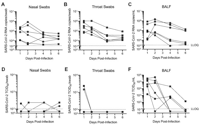
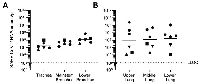
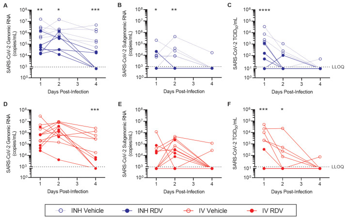
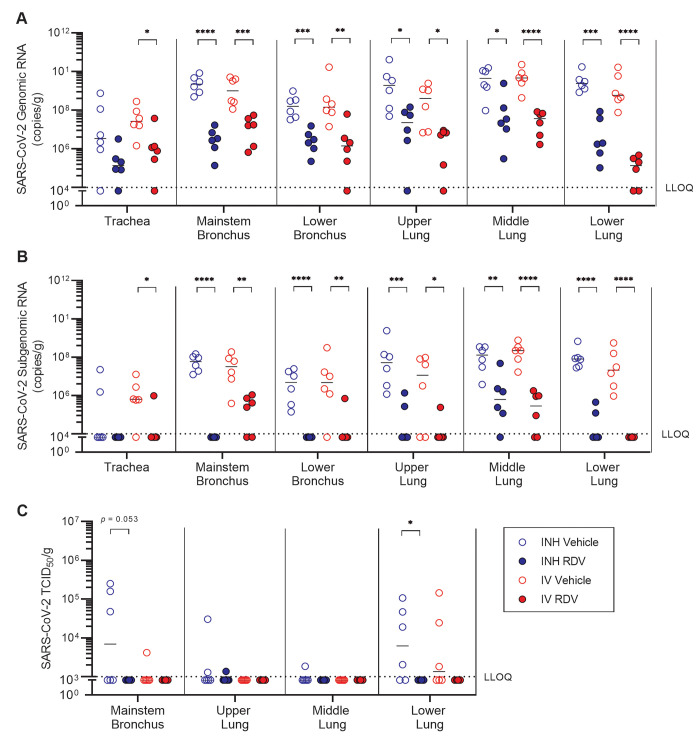
Remdesivir (RDV) is a nucleotide analog prodrug with demonstrated clinical benefit in patients with coronavirus disease 2019 (COVID-19). In October 2020, the US FDA approved intravenous (IV) RDV as the first treatment for hospitalized COVID-19 patients. Furthermore, RDV has been approved or authorized for emergency use in more than 50 countries. To make RDV more convenient for non-hospitalized patients earlier in disease, alternative routes of administration are being evaluated. Here, we investigated the pharmacokinetics and efficacy of RDV administered by head dome inhalation in African green monkeys (AGM). Relative to an IV administration of RDV at 10 mg/kg, an approximately 20-fold lower dose administered by inhalation produced comparable concentrations of the pharmacologically active triphosphate in lower respiratory tract tissues. Distribution of the active triphosphate into the upper respiratory tract was also observed following inhaled RDV exposure. Inhalation RDV dosing resulted in lower systemic exposures to RDV and its metabolites as compared with IV RDV dosing. An efficacy study with repeated dosing of inhaled RDV in an AGM model of SARS-CoV-2 infection demonstrated reductions in viral replication in bronchoalveolar lavage fluid and respiratory tract tissues compared with placebo. Efficacy was observed with inhaled RDV administered once daily at a pulmonary deposited dose of 0.35 mg/kg beginning approximately 8 hours post-infection. Moreover, the efficacy of inhaled RDV was similar to that of IV RDV administered once at 10 mg/kg followed by 5 mg/kg daily in the same study. Together, these findings support further clinical development of inhalation RDV.


Figures

Fig. 1.
Fig. 1.. Inhaled remdesivir (RDV) results in similar RDV-TP concentrations in the respiratory tract despite lower systemic and non-respiratory exposures.
Plasma concentrations of (A) RDV and its (B) alanine metabolite and (C) nucleoside metabolite were determined after a single dose of RDV administered by intravenous (IV, 10 mg/kg) or inhalation (0.17 or 0.54 mg/kg deposited dose) routes to AGM (n=3 to 4 per treatment group). Tissue concentrations of the active triphosphate metabolite RDV-TP were measured in (D) respiratory tissues, (E) liver and kidney, (F) and PBMCs approximately 24 hours post-exposure. Values are presented as mean ± SD. ns: not significant, **p <0.01, ****p <0.0001; data were analyzed using a repeated measures two-way ANOVA with Bonferroni post-hoc correction.
Fig. 2.
Fig. 2.. SARS-CoV-2 replicates in both the upper and lower respiratory tract of AGM.
AGM (n=6) were infected with SARS-CoV-2 by intranasal and intratracheal instillation. SARS-CoV-2 (A to C) RNA and (D to F) infectious viral titers were quantified from nasal (A, D) and throat (B, E) swabs, as well as bronchoalveolar lavage fluid (BALF) (C, F) collected through 6 days post-infection. SARS-CoV-2 RNA copies were quantified by RT-qPCR. Infectious SARS-CoV-2 titers were determined by a TCID50 assay. Each connected line represents an individual animal. LLOQ indicates lower limit of quantification.
Fig. 3.
Fig. 3.. SARS-CoV-2 infection is evenly distributed across the conducting airways and lung lobes collected six days post-infection.
AGM (n=6) were infected with SARS-CoV-2 by intranasal and intratracheal instillation. SARS-CoV-2 RNA was quantified in (A) conducting airways and (B) lung tissue at 6 days post-infection by RT-qPCR. Individual animals are represented by unique symbols. Horizontal bars represent the mean value for each tissue. LLOQ indicates lower limit of quantification.
Fig. 4.
Fig. 4.. Treatment with inhaled RDV following SARS-CoV-2 infection reduces viral load in bronchoalveolar lavage fluid.
AGM were inoculated with SARS-CoV-2 and treated with (A to C) inhaled or (D to F) IV RDV (closed symbols) or vehicle (open symbols) beginning 8 hours post-infection (n=6 per group). Bronchoalveolar lavage fluid was collected 1, 2 and 4 days post-inoculation. SARS-CoV-2 genomic (A, D) and subgenomic (B, E) RNA copies were quantified by RT-qPCR. Infectious SARS-CoV-2 titers (C, F) were determined by a TCID50 assay. Samples that were below the lower limit of quantification (LLOQ, dotted lines) for the assay were assigned the LLOQ for analyses. *p <0.05, **p <0.01, ***p <0.001, ****p <0.0001; data were analyzed by a repeated measures two-way ANOVA with Bonferroni post-hoc correction.
Fig. 5.
Fig. 5.. Treatment with inhaled RDV following SARS-CoV-2 infection reduces viral load in respiratory tract tissues.
AGM were inoculated with SARS-CoV-2 and treated with inhaled (blue symbols) or IV (red symbols) RDV (closed symbols) or vehicle (open symbols) beginning at 8 hours post-infection (n=6 each). Respiratory tissues were collected 6 days post-inoculation. SARS-CoV-2 (A) genomic and (B) subgenomic RNA copies were quantified by RT-qPCR. (C) Infectious SARS-CoV-2 titers were determined by a TCID50 assay. Samples that were below the lower limit of quantification (LLOQ, dotted lines) for the assay were assigned a value equal to the LLOQ for analyses. Horizontal bars indicate median values. *p <0.05, **p <0.01, ***p <0.001, ****p <0.0001; data were analyzed by a repeated measures two-way ANOVA with Bonferroni post-hoc correction.

References

    1. de Wit E., van Doremalen N., Falzarano D., Munster V. J., SARS and MERS: Recent insights into emerging coronaviruses. Nat. Rev. Microbiol. 14, 523–534 (2016). 10.1038/nrmicro.2016.81 - DOI - PMC - PubMed
    1. Ksiazek T. G., Erdman D., Goldsmith C. S., Zaki S. R., Peret T., Emery S., Tong S., Urbani C., Comer J. A., Lim W., Rollin P. E., Dowell S. F., Ling A.-E., Humphrey C. D., Shieh W.-J., Guarner J., Paddock C. D., Rota P., Fields B., DeRisi J., Yang J.-Y., Cox N., Hughes J. M., LeDuc J. W., Bellini W. J., Anderson L. J.; SARS Working Group , A novel coronavirus associated with severe acute respiratory syndrome. N. Engl. J. Med. 348, 1953–1966 (2003). 10.1056/NEJMoa030781 - DOI - PubMed
    1. Assiri A., Al-Tawfiq J. A., Al-Rabeeah A. A., Al-Rabiah F. A., Al-Hajjar S., Al-Barrak A., Flemban H., Al-Nassir W. N., Balkhy H. H., Al-Hakeem R. F., Makhdoom H. Q., Zumla A. I., Memish Z. A., Epidemiological, demographic, and clinical characteristics of 47 cases of Middle East respiratory syndrome coronavirus disease from Saudi Arabia: A descriptive study. Lancet Infect. Dis. 13, 752–761 (2013). 10.1016/S1473-3099(13)70204-4 - DOI - PMC - PubMed
    1. Choi W. S., Kang C. I., Kim Y., Choi J. P., Joh J. S., Shin H. S., Kim G., Peck K. R., Chung D. R., Kim H. O., Song S. H., Kim Y. R., Sohn K. M., Jung Y., Bang J. H., Kim N. J., Lee K. S., Jeong H. W., Rhee J.-Y., Kim E. S., Woo H., Oh W. S., Huh K., Lee Y. H., Song J. Y., Lee J., Lee C.-S., Kim B.-N., Choi Y. H., Jeong S. J., Lee J.-S., Yoon J. H., Wi Y. M., Joung M. K., Park S. Y., Lee S. H., Jung S.-I., Kim S.-W., Lee J. H., Lee H., Ki H. K., Kim Y.-S.; Korean Society of Infectious Diseases , Clinical Presentation and Outcomes of Middle East Respiratory Syndrome in the Republic of Korea. Infect. Chemother. 48, 118–126 (2016). 10.3947/ic.2016.48.2.118 - DOI - PMC - PubMed
    1. Who Mers-Cov Research Group , State of Knowledge and Data Gaps of Middle East Respiratory Syndrome Coronavirus (MERS-CoV) in Humans. PLOS Curr. 5, ••• (2013). - PMC - PubMed

Publication types

LinkOut - more resources