

Review
doi: 10.1016/j.urology.2021.12.013.
Epub 2021 Dec 27.
Bilateral Renal Mucor Mycosis Presenting as Anuria in a Covid 19 Recovered Patient: A Case Report and Review of Literature
Affiliations
- PMID: 34968570
- PMCID: PMC8711140
- DOI: 10.1016/j.urology.2021.12.013
Item in Clipboard
Review
Bilateral Renal Mucor Mycosis Presenting as Anuria in a Covid 19 Recovered Patient: A Case Report and Review of Literature
Urology.
2022 Mar.
No abstract available
Figures
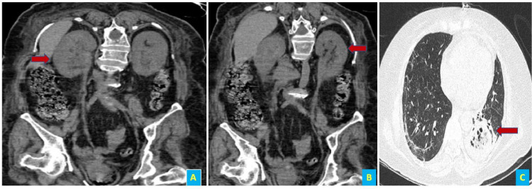
(A,B). NCCT abdomen showing bilateral perinephric fat stranding and bilateral mild hydroureteronephrosis.(red arrows) (C). CT Chest showing peripheral patchy interstitial thickening with fibrotic bands with organizing pneumonia changes in left lower lobe.(red arrows). (Color version available online.)

RGP showing bilateral multiple filling defects involving ureter, renal pelvis and calyces (A and C). (B) Fluoroscopic image of right PCN insertion. (D) Fluoroscopic image showing left double J stent placement. (Color version available online.)
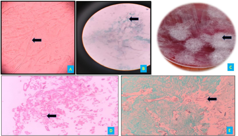
Microbiological diagnosis of Mucormycosis.(A-C) (40x magnification) (A) KOH preparation showing aseptate broad hyaline hyphal filaments with wide angle branching(Day 1). (B). GMS(Gomori's methenamine silver stain (GMS) stain showing black aseptate hyphal forms. (C) Growth on blood agar showing rapidly growing mycelial colonies having hairy appearance - lid lifting sign. Histopathological diagnosis of Mucormycosis. (D and E) H&E stain (D) and GMS stain (E) at 40x magnification: Numerous collapsible aseptate non branching fungal hyphae (D&E) in necro inflammatory debris (D) (black arrow). (Color version available online.)
References
-
- Cascella M, Rajnik M, Aleem A, et al. StatPearls [Internet] StatPearls Publishing; Treasure Island (FL): 2021. Features, evaluation, and treatment of coronavirus (COVID-19) [Updated 2021 Apr 20]https://www.ncbi.nlm.nih.gov/books/NBK554776/ Available from: - PubMed
-
- Indian Council of Medical Research. Evidence based advisory in the time of COVID-19 (screening, diagnosis & management of mucormycosis). May 9, 2021.https://www.icmr.gov.in/pdf/covid/techdoc/Mucormycosis_ADVISORY_FROM_ICM....
Publication types
MeSH terms
LinkOut - more resources
Full Text Sources
Medical
