. 2022 Feb;18(2):216-225.
doi: 10.1038/s41589-021-00932-1. Epub 2021 Dec 30.

Engineering CAR-T cells to activate small-molecule drugs in situ

Affiliations

Engineering CAR-T cells to activate small-molecule drugs in situ

Thomas J Gardner et al. Nat Chem Biol. 2022 Feb.

Abstract

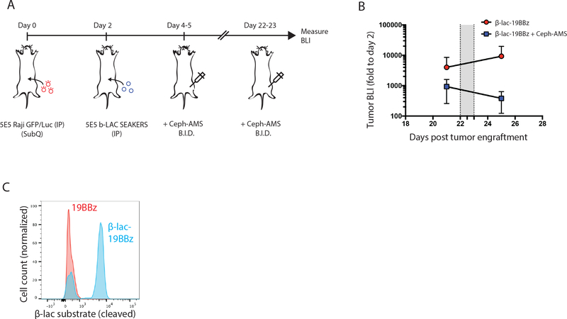
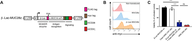
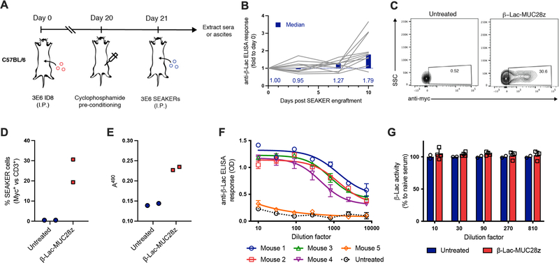
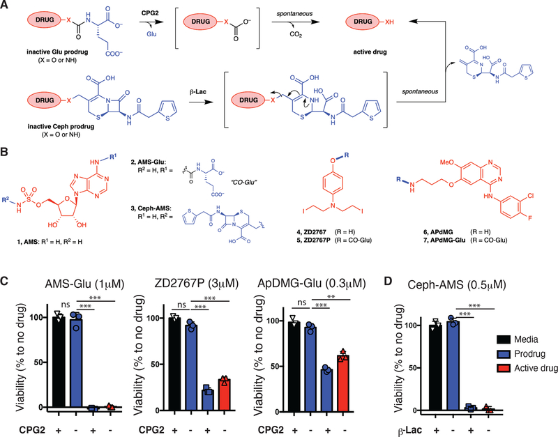
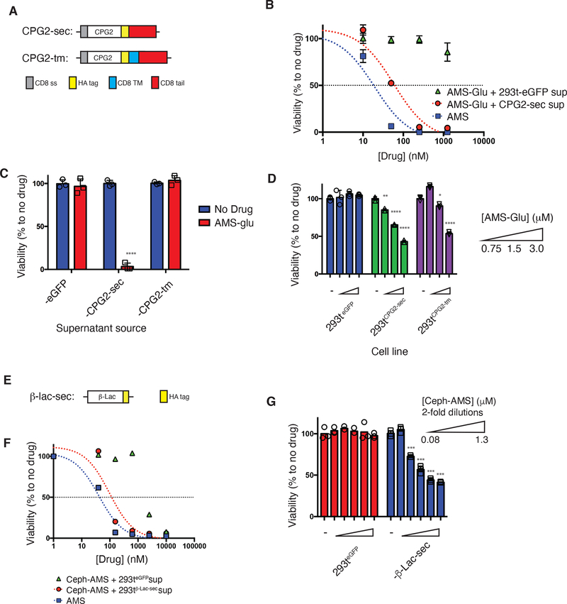
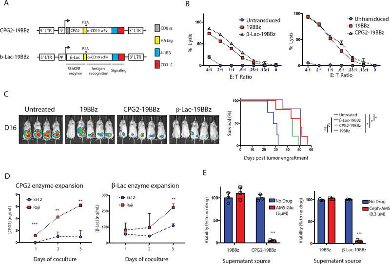
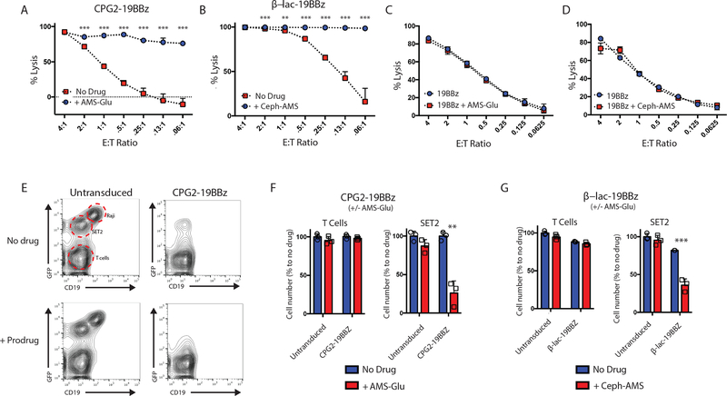
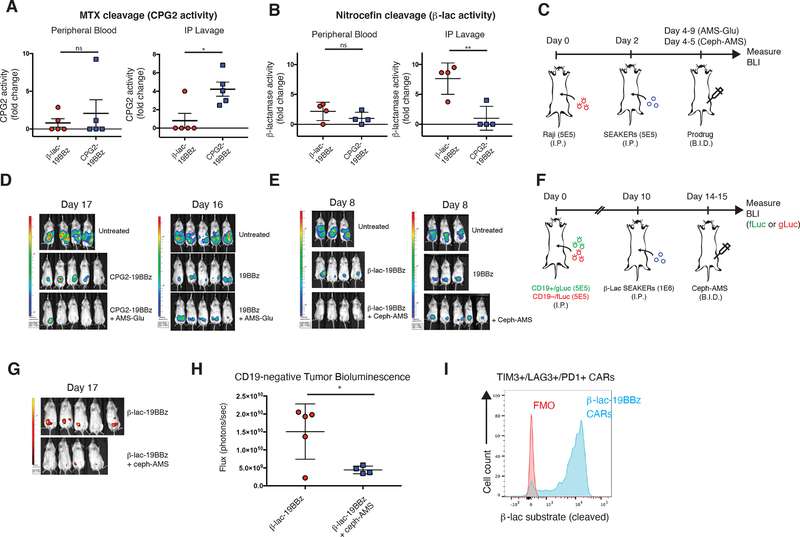
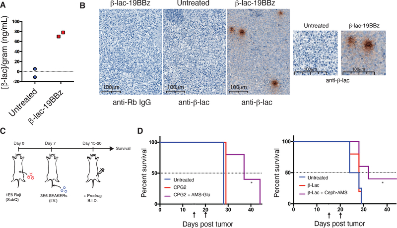
Chimeric antigen receptor (CAR)-T cells represent a major breakthrough in cancer therapy, wherein a patient's own T cells are engineered to recognize a tumor antigen, resulting in activation of a local cytotoxic immune response. However, CAR-T cell therapies are currently limited to the treatment of B cell cancers and their effectiveness is hindered by resistance from antigen-negative tumor cells, immunosuppression in the tumor microenvironment, eventual exhaustion of T cell immunologic functions and frequent severe toxicities. To overcome these problems, we have developed a novel class of CAR-T cells engineered to express an enzyme that activates a systemically administered small-molecule prodrug in situ at a tumor site. We show that these synthetic enzyme-armed killer (SEAKER) cells exhibit enhanced anticancer activity with small-molecule prodrugs, both in vitro and in vivo in mouse tumor models. This modular platform enables combined targeting of cellular and small-molecule therapies to treat cancers and potentially a variety of other diseases.


Figures

Extended Data Fig. 1
Extended Data Fig. 1. Synthesis of AMS-Glu prodrug (2).
Synthesis of AMS-Glu prodrug (2). DMA = N,N-dimethylacetamide; DMF = N,N-dimethylformamide; TBAF = tetrabutylammonium fluoride; TBS = t-butyldimethylsilyl.
Extended Data Fig. 2
Extended Data Fig. 2. Synthesis of Ceph-AMS prodrug (3).
Synthesis of Ceph-AMS prodrug (3). (a) Synthesis of protected AMS precursor S9. (b) Synthesis of Ceph-AMS (3). pyr = pyridine; TFA = trifluoroacetic acid; THF = tetrahydrofuran; TMS = trimethylsilyl.
Extended Data Fig. 3
Extended Data Fig. 3. Complete bioluminescent imaging data for in vivo efficacy in mouse intraperitoneal Raji tumor xenografts
Complete bioluminescent imaging data for in vivo efficacy in mouse intraperitoneal Raji tumor xenografts treated with (a) CPG2–19BBz SEAKER cells and AMS-Glu (2) (50 mg/kg, ip, bid, days 2–7 post-CAR engraftment, 12 doses total, gray band) or (b) β-Lac-19BBz SEAKER cells and Ceph-AMS (3) (4 mg/kg, ip, bid, days 2–3 post CAR engraftment, 3 doses total, gray band). Raw BLI is plotted on log scale; AUC is plotted on split linear scale. (mean ± s.d. of n = 5 mice per group; experiment was repeated with similar results.) Representative images are shown in Fig. 5d and 5e, respectively, of the manuscript.
Extended Data Fig. 4
Extended Data Fig. 4. Complete bioluminescent imaging data for in vivo efficacy against antigen-negative cells in intraperitoneal heterogeneous tumor xenografts.
Complete bioluminescent imaging data for in vivo efficacy against antigen-negative cells in intraperitoneal heterogeneous tumor xenografts. (a) Bioluminescent imaging (BLI) and quantification of CD19+ Nalm6 cells expressing mCherry and Gaussia luciferase (Nalm6-mCherry/gLuc [CD19+]) in untreated mice or mice receiving β-Lac-19BBz SEAKER cells plus or minus 3 injections of Ceph-AMS (3: 4 mg/kg, ip, bid). Images taken at day 20 post tumor engraftment; one mouse in the group treated with SEAKER cells and prodrug showed tumor clearance but died after day 14 and is omitted. Complete radiance data is shown in the right two panels, with raw BLI plotted on log scale and AUC plotted on a linear scale. (Mean ± s.d. of n = 4 (untreated, SEAKER+prodrug) or n = 5 (SEAKER alone) mice per group; Student’s two-tailed t-test: ns = not significant; *p<0.05; experiment was performed once). (b) BLI and quantification of CD19– Nalm6 cells expressing eGFP and firefly luciferase (Nalm6-eGFP/fLuc [CD19–]) in untreated mice or mice receiving β-Lac-19BBz SEAKER cells plus or minus 3 injections of Ceph-AMS (3: 4 mg/kg, ip, bid). Images taken at day 17 post tumor engraftment; one mouse in the group treated with SEAKER cells and prodrug died after day 14 and is omitted. The left two panels also appear in Fig. 5g,h of the manuscript. Complete radiance data is shown in the right two panels, with raw BLI plotted on log scale and AUC plotted on a linear scale. (Mean ± s.d. of n = 4 (untreated, SEAKER+prodrug) or n = 5 (SEAKER alone) mice per group; Student’s two-tailed t-test: ns = not significant; *p<0.05; experiment was performed once).
Extended Data Fig. 5
Extended Data Fig. 5. Persistence of β-Lacenzyme activity and Ceph-AMS prodrug activation in mouse intraperitoneal Raji tumor xenografts after SEAKER cell exhaustion in vivo.
Persistence of β-Lac enzyme activity and Ceph-AMS prodrug activation in mouse intraperitoneal Raji tumor xenografts after SEAKER cell exhaustion in vivo. (a) Experimental scheme for rescue of relapsed Raji xenograft by treatment with Ceph-AMS (3: 4 mg/kg, ip, bid, days 4–5, 3 doses total, then days 22–23 (gray bar), 3 doses total) after exhaustion of β-Lac-19BBz SEAKER cells. (b) Quantitation of tumor bioluminescence before and after second dosing period (median with s.d. of n = 4 mice per group; experiment was performed once). (c) Persistence of β-Lac enzyme activity in β-Lac-19BBz SEAKER cells extracted from two of the mice from the experiment in panel a at day 30 (day 28 post administration), in comparison to standard 19BBz CAR-T cell controls (flow cytometry analysis: β-Lac substrate = CCF2-AM).
Extended Data Fig. 6
Extended Data Fig. 6. Construction and characterization of βLac-expressing murine SEAKER cells.
Construction and characterization of β-Lac-expressing murine SEAKER cells. (a) SEAKER construct encoding secreted β-Lac and a murine CAR: β-Lac-MUC28z (β-Lac/α-MUC16/CD28/CD3ζ). LTR = long terminal, Ψ = psi packaging element, FLAG = FLAG epitope tag (pink), P2A = 2A self cleaving peptide, α-MUC16 scFv = MUC-16-specific mouse-derived single chain variable fragment, myc = Myc epitope tag (brown), mCD28 = mouse CD28 costimulatory domain (green), mCD3ζ = mouse CD3 zeta chain (red). (b) Flow cytometry analysis of α-MUC16 CAR expression in retrovirally-transduced primary mouse T cells (fluorescently (phycoerythrin, PE) labeled anti-idiotype antibody; representative data from 5 independent experiments). (c) Trans-cytotoxicity of supernatant fluid (sn) from β-Lac-MUC28z SEAKER cells with or without Ceph-AMS (3: 107 nM) against mouse EL4 lymphoma cells, compared to prodrug alone and parent drug AMS (1: 500 nM) (24 h, CellTiter-Glo assay; mean ± s.d. of n = 3 technical replicates/samples; Student’s two-tailed t-test: ns = not significant, ***p<0.001; experiment was perfomed once).
Extended Data Fig. 7
Extended Data Fig. 7. Assessment of SEAKER cell immunogenicity in an immunocompetent mouse model.
Assessment of SEAKER cell immunogenicity in an immunocompetent mouse model. (a) Experimental scheme to assess immunogenicity of β-Lac-MUC28z SEAKER cells in a syngeneic intraperitoneal ID8 tumor model. Sera were collected on Days 21, 24, 28, and 31 and tested for anti-β-Lac antibodies in panel b. In a separate experiment, ascites were recovered on Day 28 by peritoneal lavage and tested for the presence of SEAKER cells in panels c,d and β-Lac enzyme activity in panel e. (b) Detection anti-β-Lac antibodies in sera over 10 days following SEAKER cell engraftment (Days 21–31) (median with lines representing each individual mouse of n = 12; experiment was performed once). (c,d) Flow cytometry analysis of SEAKER cell (myc+) persistence among T cells (CD3+) and (e) nitrocefin cleavage-based quantitation of β-Lac enzyme activity in ascites recovered 7 days after SEAKER cell engraftment (Day 28) (representative data shown from n = 2 mice per group; on average, 25% of T cells were SEAKER-positive; experiment was performed once). SSC = side scatter. (f) In a third experiment, mice were treated as in panel a, but without cyclophosphamide pretreatment to maximize the antibody response, then sera were recovered 5 days after SEAKER cell engraftment (Day 26) and analyzed for anti-β-Lac antibodies (n = 1 mouse in untreated group; n = 5 mice in treated group; mean ± s.d. of n = 3 technical replicates from each mouse; experiment was performed once). Sera from the 4 mice showing anti-β-Lac antibodies were used for the ex vivo enzyme activity experiment in panel g. (g) Nitrocefin cleavage-based quantitation of enzyme activity of recombinant β-Lac treated with sera from untreated or SEAKER-treated mice from panel f (n = 2 mice in untreated group; mean ± s.d. of n = 4 mice in treated group; experiment was performed once).
Figure 1.
Figure 1.. Modular prodrug designs for use with SEAKER cells.
(a) Glutamate-masked prodrugs can be cleaved with Pseudomonas sp. CPG2 to form a carbonic acid intermediate, followed by spontaneous decomposition of the linker to form the active drug. Cephalothin-masked prodrugs can be cleaved by Enterobacter cloacae β-Lac to form a hydrolyzed intermediate, followed by spontaneous elimination of the cephalothin byproduct to form the active drug. Drugs are shown in red, masks in blue, linkers in gray. (b) Structures of cytotoxic natural product AMS (1), glutamate-masked prodrug AMS-Glu (2), and cephalothin-masked prodrug Ceph-AMS (3); nitrogen mustard ZD2767 (4) and glutamate-masked prodrug ZD2767P (5); and targeted kinase inhibitor APdMG (6) and glutamate-masked prodrug APdMG-Glu (7). (AMS = adenosine-5´-O-monosulfamate or 5´-O-sulfamoyladenosine; APdMG = 7-O-aminopropyl-7-O-des[morpholinopropyl]gefitinib). (c) Cytotoxicity of prodrug AMS-Glu (2: 1 μM), ZD2767P (5: 3 μM), APdMG-Glu (7: 0.3 μM) with or without recombinant CPG2 (250 ng/mL), or of the parent drug AMS (1), ZD2767 (4), APdMG (6), to SET2 cells (48 h, CellTitre-Glo assay). (d) Cytotoxicity of prodrug Ceph-AMS (3: 0.5 μM), with or without recombinant β-Lac (10 ng/mL), or of the parent drug AMS (1: 0.5 μM) to SET2 cells (48 h, CellTitre-Glo assay). (For c,d: mean ± s.d. of n = 3 technical replicates/samples; Student’s two-tailed t-test: ns = not significant, **p<0.01, ***p<0.001; representative of 2 or more experiments.)
Figure 2.
Figure 2.. In vitro validation of prodrug activation by CPG2- and β-Lac-expressing HEK293T cells.
(a) CPG2 gene cassettes generated for eukaryotic expression: CPG2-sec (secreted), CPG2-tm (membrane-anchored). CD8 ss = CD8 signal peptide (gray), CPG2 = native CPG2 gene excluding endogenous signal peptide (aa1–22) (white), HA tag = hemagglutinin epitope tag (yellow), CD8 TM = CD8 transmembrane domain (blue), CD8 tail = CD8 cytosolic tail (red). (b) Trans-cytotoxicity of supernatant fluids from HEK293T-CPG2-sec cells (red circles) or control cells (green triangles) with increasing concentrations of prodrug AMS-Glu (2), or of parent drug AMS (1) (blue squares) against SET2 target cells (48 h, CellTiter-Glo assay). (c) Trans-cytotoxicity of supernatant fluids from HEK293T-eGFP, -CPG2-sec, and -CPG2-tm cell lines with or without AMS-Glu (2: 5 μM) against SET2 target cells (48 h, CellTiter-Glo assay). (d) Cis-cytotoxicity of increasing concentrations of AMS-Glu (2) to HEK293T-eGFP, -CPG2-sec, and -CPG2-tm cell lines (96 h, CellTitre-Glo assay). (e) β-Lac-sec (secreted) gene cassette generated for eukaryotic expression: β-Lac = native β-Lac gene excluding endogenous signal peptide (aa1–20) (white), HA tag = hemagglutinin epitope tag (yellow). (f) Trans-cytotoxicity (against other cell lines) of supernatant fluids from HEK293T-β-Lac-sec cells (red circles) or control cells (green triangles) with increasing concentration of prodrug Ceph-AMS (3), or of parent drug AMS (1) (blue squares) against SET2 antigen-negative target cells (48 h, CellTiter-Glo). (g) Cis-cytotoxicity (self-killing) of increasing concentrations of Ceph-AMS (3) to HEK293T-eGFP and HEK293T-β-Lac-sec cell lines (48 h, CellTitre-Glo). (For b-d; f-g: mean ± s.d. of n = 3 biological replicates/samples; Student’s two-tailed t-test: *p<0.05, **p<0.01, ***p<0.001, ****p<0.0001; representative of 2 or more experiments.)
Figure 3.
Figure 3.. Construction and characterization of CPG2- and β-Lac-expressing SEAKER cells.
(a) SEAKER CAR constructs encoding secreted prodrug-activating enzymes: CPG2–19BBz (CPG2/α-CD19/4–1BB/CD3ζ) and β-Lac-19BBz (β-Lac/α-CD19/4–1BB/CD3ζ). LTR = long terminal repeat, Ψ = psi packaging element, CD8 ss = CD8 signal peptide (gray), HA tag = hemagglutinin epitope tag (yellow), P2A = 2A self cleaving peptide, α-CD19 scFv = CD19-specific single chain variable fragment, 4–1BB = 4–1BB costimulatory domain (blue), CD3ζ = CD3 zeta chain (red). (b) Cytolytic activity of standard 19BBz CAR-T cells, CPG2–19BBz SEAKER cells, and β-Lac-19BBz SEAKER cells against Raji (CD19+) target cells expressing firefly luciferase (18 h, bioluminescence assay; mean ± s.d. of n = 3 biological replicates/sample; representative data from 3 independent donors). (c) Antitumor efficacy of standard 19BBz CAR-T cells, CPG2–19BBz SEAKER cells, and β-Lac-19BBz SEAKER cells, without prodrugs, against Raji xenografts in NSG mice (day 16 post-tumor engraftment, bioluminescent imaging (left) and Kaplan–Meier curve (right). (Log-rank (Mantel–Cox) test: *p<0.05; untreated vs. β-Lac-19BBz: p = 0.003; untreated vs. CPG2–19BBz: p = 0.023; Untreated vs. 19BBz: p = 0.03. Experiment was performed once.) (d) SEAKER enzyme expression in cocultures of anti-CD19 SEAKERS with Raji (CD19+) or SET2 (CD19−) cells (CPG2: ELISA assay; β-Lac: nitrocefin cleavage UV assay; mean ± s.d. of n = 3 biological replicates/samples; Student’s two-tailed t-test: **p<0.01, ***p<0.001; representative of 3 experiments). (e) Trans-cytotoxicity of supernatant fluids from standard 19BBz CAR-T cells and SEAKER cells, with or without the corresponding prodrug, against SET2 target cells (48 h, CellTiter-Glo) (mean ± s.d. of n = 3 biological replicates/samples; Student’s two-tailed t-test: **p<0.01, ***p<0.001).
Figure 4.
Figure 4.. In vitro validation of prodrug activation by SEAKER cells and antigen-negative cell killing.
(a) Specific lysis by CPG2–19BBz SEAKER cells, with and without AMS-Glu (2: 20 μM), against Raji (CD19+) target cells expressing firefly luciferase (18–96 h, bioluminescence assay). (b) Specific lysis of β-Lac-19BBz SEAKER cells, with and without Ceph-AMS (3: 0.3 μM), as in panel a. Specific lysis by 19BBz CAR-T cells, with and without AMS-Glu (2) (c) or Ceph-AMS (3) (d) against Raji (CD19+) target cells expressing firefly luciferase (48 h, bioluminescence assay). (e) Flow cytometric analysis of trans-cytotoxicity of CPG2–19BBz SEAKER cells, with and without AMS-Glu (2: 20 μM), against Raji (CD19+) antigen-positive cells and SET2 (CD19−) antigen-negative cells engineered to express eGFP (representative data from 2 donors). (f) Quantitation of cell numbers from coculture experiments in panel e. (g) Quantitation of cell numbers from flow cytometric analysis of trans-cytotoxicity of β-Lac-19BBz SEAKER cells, with and without Ceph-AMS (3: 0.3 μM) against Raji (CD19+) antigen-positive cells and SET2 (CD19−) antigen-negative cells. (For a-g: mean ± s.d. of n = 3 biological replicates; Student’s two-tailed t-test: **p<0.01, ***p<0.001; representative data from 3 or more independent donors.)
Figure 5.
Figure 5.. Enhanced in vivo efficacy of prodrug–SEAKER cell combinations in mouse intraperitoneal xenografts.
(a) CPG2 enzyme activity in peripheral blood (left) or peritoneal lavage (right) of Raji tumor-engrafted NSG mice treated with CPG2–19BBz SEAKER cells or β-Lac-19BBz SEAKER cells (negative control), based on methotrexate cleavage assay (fold-change; mean ± s.d. of n = 5 mice per group; Student’s two-tailed t-test: *p<0.05; representative data from 2 independent experiments). (b) β-Lac enzyme activity in peripheral blood (left) or peritoneal lavage (right) of Raji tumor-engrafted mice treated with CPG2–19BBz SEAKER cells (negative control) or β-Lac-19BBz SEAKER cells, based on nitrocefin cleavage assay (fold-change; mean ± s.d. of n = 4 mice per group; Student’s two-tailed t-test: **p<0.01; representative data from 2 independent experiments). (c) Experimental scheme to assess efficacy of SEAKER–prodrug combinations in intraperitoneal tumor model (AMS-Glu (2): 50 mg/kg, ip, bid, 12 doses total; Ceph-AMS (3): 4 mg/kg, ip, bid, 3 doses total). (d,e) Tumor bioluminescence was monitored over time (representative images shown; experiment in panel e was repeated with similar results) (see also Extended Data Fig. 3). (f) Experimental scheme to assess efficacy in a heterogeneous tumor model (2 × 10⁶ total cells, 1:1 Nalm6-mCherry/gLuc (CD19+) and Nalm6-eGFP/fLuc (CD19−). (Ceph-AMS (3): 4 mg/kg, ip, bid, 3 doses total). (g,h) Antitumor efficacy against CD19− Nalm6 cancer cells (fLuc) engrafted within the heterogeneous tumor with CD19+ Nalm6 cancer cells (see also Extended Data Fig. 4), only in mice receiving β-Lac-19BBz SEAKER cells plus Ceph-AMS prodrug (3) (representative images shown on day 17 from 20-day study; one mouse in prodrug-treated group died between day 14 and day 17 and is omitted) (mean ± s.d. of n = 5 mice in SEAKER-treated group and n = 4 mice in SEAKER+prodrug-treated group; Student’s two-tailed t-test: *p<0.05; experiment was performed once). (i) Retention of β-Lac enzyme activity in β-Lac-19BBz SEAKER cells expressing T-cell exhaustion markers (TIM3, LAG3, PD1) after 26 days in Raji-engrafted mice (flow cytometry analysis: FMO = fluorescence minus one control; β-Lac substrate = CCF2-AM) (representative data shown for one of n = 4 mice; representative data from 2 independent experiments).
Figure 6.
Figure 6.. Enhanced in vivo efficacy of Ceph-AMS–β-Lac-SEAKER cell combinations in mouse subcutaneous xenografts.
(a) Nitrocefin cleavage-based quantitation of tumor β-Lac concentration in a subcutaneous Raji tumor from a mouse treated with 3 × 10⁶ β-Lac-19BBz SEAKER cells (IV) (representative data from two of n = 5 mice per group in 3 independent experiments). (b) anti-β-Lac immunohistochemistry imaging of subcutaneous Raji tumors extracted on day 15 from mice that were untreated, or received 3 × 10⁶ β-Lac-19BBz SEAKER cells (IV) (top panels – left: isotype control; center: untreated mouse stained with anti-β-Lac antibody; right: β-Lac-19BBz-treated mouse stained with anti-β-Lac antibody). Increased magnification highlights diffuse β-Lac staining throughout the tumor environment (bottom panels) (representative data shown from one of the 3 remaining mice from the n = 5 group used in panel a). (c) Experimental scheme to assess therapeutic efficacy of SEAKER–prodrug combinations in a subcutaneous solid tumor model. Raji tumor cells were engrafted sq on day 0 followed by SEAKER cells iv on day 7. The corresponding prodrug was administered beginning on day 15 (AMS-Glu (2): 50 mg/kg, ip, bid, days 15–20, 12 doses total, or Ceph-AMS (3): 4 mg/kg, ip, bid every other day, days 15, 17, 19, 6 doses total) and mice were monitored for survival. (d) Survival analysis of mice engrafted with subcutaneous Raji tumors receiving subtherapeutic doses of CPG2–19BBz SEAKER cells plus AMS-Glu (2) (left panel), or β-Lac-19BBz SEAKER cells plus Ceph-AMS (3) (right panel). (Arrows denote beginning and end of the prodrug administration period; n = 5 mice/group; log-rank (Mantel–Cox) test: *p<0.05; CPG2–19BBz vs. CPG2–19BBz + AMS-Glu: p = 0.023; β-Lac-19BBz vs. β-Lac-19BBz + Ceph-AMS: p = 0.048; experiment was repeated with similar results).

Comment in

References

    1. Feldman SA, Assadipour Y, Kriley I, Goff SL & Rosenberg SA Adoptive cell therapy—Tumor-infiltrating lymphocytes, T-cell receptors, and chimeric antigen receptors. Semin. Oncol. 42, 626–639 (2015). - PMC - PubMed
    1. Leon E, Ranganathan R & Savoldo B Adoptive T cell therapy: Boosting the immune system to fight cancer. Semin. Immunol. 49, 101437 (2020). - PMC - PubMed
    1. Sadelain M, Rivière I & Riddell S Therapeutic T cell engineering. Nature 545, 423–431 (2017). - PMC - PubMed
    1. Beyar-Katz O & Gill S Advances in chimeric antigen receptor T cells. Curr. Opin. Hematol. 27, 368–377 (2020). - PubMed
    1. June CH, O’Connor RS, Kawalekar OU, Ghassemi S & Milone MC CAR T cell immunotherapy for human cancer. Science 359, 1361–1365 (2018). - PubMed

Publication types