

Imaging findings in invasive rhino-orbito-cerebral mucormycosis in post-COVID-19 patients
- PMID: 34970028
- PMCID: PMC8682837
- DOI: 10.1080/08998280.2021.1981100
Imaging findings in invasive rhino-orbito-cerebral mucormycosis in post-COVID-19 patients
Abstract
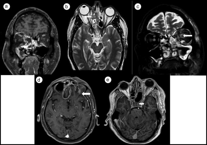
Rhino-orbito-cerebral mucormycosis (ROCM) is a life-threatening addition to the COVID-19 disease spectrum and is caused by an angioinvasive saprophytic opportunistic fungus. Early diagnosis is important to avoid disease spread and mortality. Contrast-enhanced magnetic resonance imaging plays a major role in detection of intraorbital and intracranial extension. We present imaging findings of 15 patients with post-COVID-19 rhino-orbito-cerebral mucormycosis who were diagnosed with invasive sinus mucormycosis at our institution and are currently undergoing treatment. All patients were diabetics, and 80% had a history of steroid intake during the course of COVID-19 treatment. There was a male preponderance (73.3%). The maxillary sinus was most commonly involved (86.7%). Orbital and intracranial invasion was seen in 73.3% and 60% of patients, respectively. The presence of retroantral, facial, infratemporal, and orbital fat stranding was an early sign of extrasinus spread. Other common sites of extrasinus involvement were the orbit and face, followed by the orbital apex, masticator space, pterygopalatine fossa, bone, skull base, cavernous sinus, brain, and internal carotid artery. In conclusion, early detection of extrasinus spread of mucormycosis by imaging is important so that aggressive treatment can be given and mortality can be reduced.
Keywords: COVID-19; extrasinus spread; mucormycosis.
Copyright © 2021 Baylor University Medical Center.
Figures
Similar articles
-
Magnetic Resonance Imaging Features of Rhino-Orbito-Cerebral Mucormycosis in Post-COVID-19 Patients: Radio-Pathological Correlation.Diagnostics (Basel). 2023 Apr 25;13(9):1546. doi: 10.3390/diagnostics13091546. Diagnostics (Basel). 2023. PMID: 37174937 Free PMC article.
-
Imaging Spectrum of Coronavirus Disease- 2019 Associated Rhino-Orbital-Cerebral Mucormycosis; From Sinonasal Inflammation to Intracranial Involvement.Acad Radiol. 2023 Sep;30(9):1904-1914. doi: 10.1016/j.acra.2022.12.011. Epub 2022 Dec 9. Acad Radiol. 2023. PMID: 36581530 Free PMC article.
-
The Radiological Spectrum of Rhino-Oculo-Cerebral Mucormycosis.Cureus. 2023 Jun 25;15(6):e40932. doi: 10.7759/cureus.40932. eCollection 2023 Jun. Cureus. 2023. PMID: 37519552 Free PMC article.
-
Magnetic resonance imaging of rhino-orbito-cerebral mucormycosis: a pictorial review.Acta Radiol. 2023 Apr;64(4):1641-1649. doi: 10.1177/02841851221132788. Epub 2022 Oct 17. Acta Radiol. 2023. PMID: 36254401 Review.
-
COVID-19-associated rhino-orbital-cerebral mucormycosis: A systematic review, meta-analysis, and meta-regression analysis.Indian J Pharmacol. 2021 Nov-Dec;53(6):499-510. doi: 10.4103/ijp.ijp_839_21. Indian J Pharmacol. 2021. PMID: 34975140 Free PMC article.
Cited by
-
Clinical Phenotypes of COVID-19 Associated Mucormycosis (CAM): A Comprehensive Review.Diagnostics (Basel). 2022 Dec 8;12(12):3092. doi: 10.3390/diagnostics12123092. Diagnostics (Basel). 2022. PMID: 36553099 Free PMC article. Review.
-
Spectrum of magnetic resonance imaging findings in post-COVID-19 patients presenting with rhino-orbito-cerebral mucormycosis in a teaching hospital in Malwa region of Punjab.J Family Med Prim Care. 2022 Dec;11(12):7788-7794. doi: 10.4103/jfmpc.jfmpc_1136_22. Epub 2023 Jan 17. J Family Med Prim Care. 2022. PMID: 36994047 Free PMC article.
-
Utility of intraoperative scoring system in rhino-orbital mucormycosis as a prognostic tool.Acta Otorhinolaryngol Ital. 2024 Oct;44(5):313-321. doi: 10.14639/0392-100X-N2705. Acta Otorhinolaryngol Ital. 2024. PMID: 39526768 Free PMC article.
-
Rhino-orbito-cerebral mucormycosis and its resurgence during COVID-19 pandemic: A review.Indian J Ophthalmol. 2023 Jan;71(1):39-56. doi: 10.4103/ijo.IJO_1219_22. Indian J Ophthalmol. 2023. PMID: 36588206 Free PMC article. Review.
References
-
- Hingad N, Kumar G, Deshmukh R.. Oral mucormycosis causing necrotizing lesion in a diabetic patient: a case report. Int J Oral Maxillofac Pathol. 2012;3(3):8–12.
-
- Therakathu J, Prabhu S, Irodi A, Sudhakar SV, Yadav VK, Rupa V.. Imaging features of rhinocerebral mucormycosis: a study of 43 patients. Egypt J Radiol Nucl Med. 2018;49(2):447–452. doi:10.1016/j.ejrnm.2018.01.001. - DOI
LinkOut - more resources
Full Text Sources
Miscellaneous