. 2021 Dec 21:2021:9247285.
doi: 10.1155/2021/9247285. eCollection 2021.

Sphingosine Kinase 1 Plays an Important Role in Atorvastatin-Mediated Anti-Inflammatory Effect against Acute Lung Injury

Affiliations

Sphingosine Kinase 1 Plays an Important Role in Atorvastatin-Mediated Anti-Inflammatory Effect against Acute Lung Injury

Lan Wu et al. Mediators Inflamm. .

Abstract

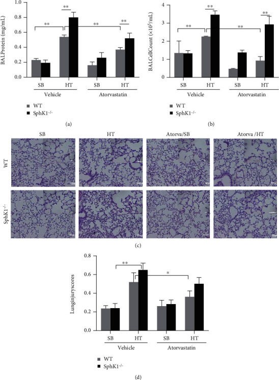
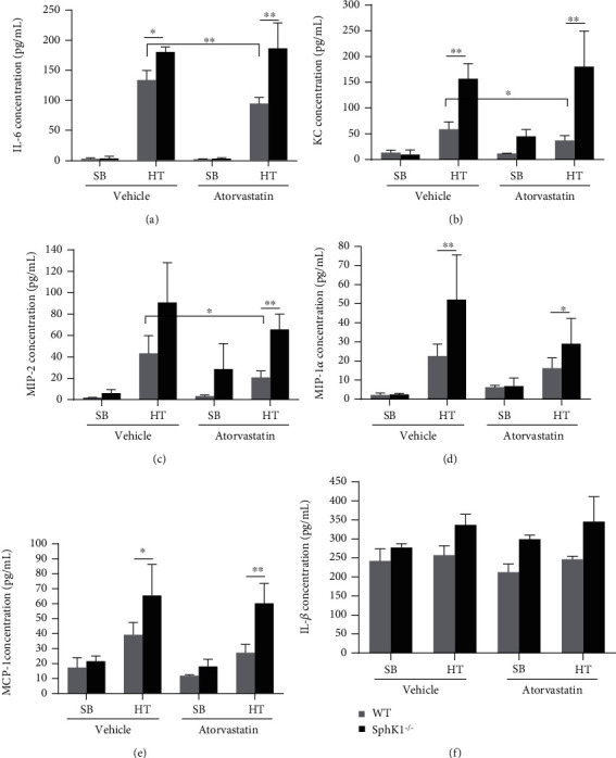
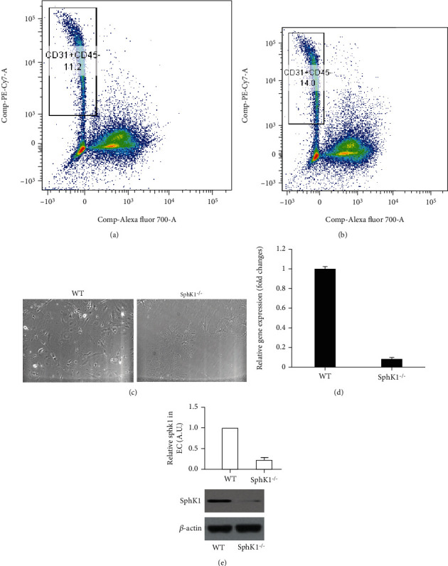
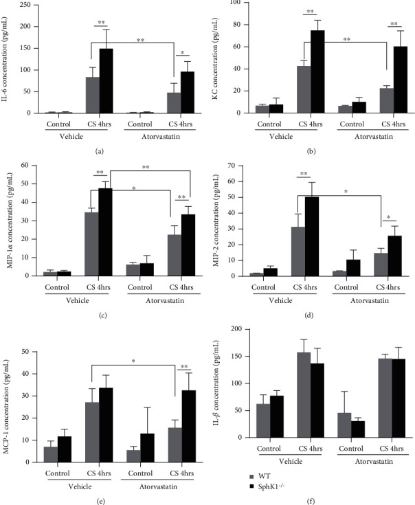
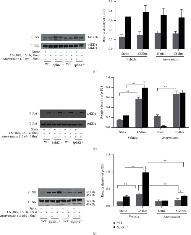
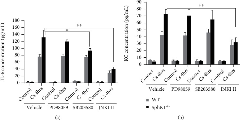
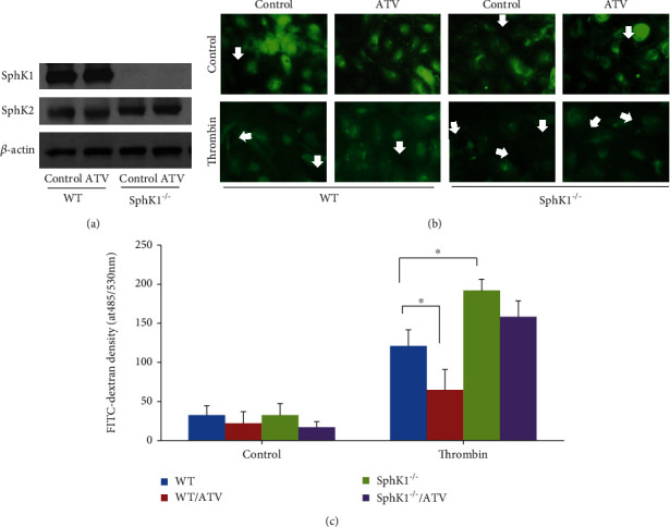
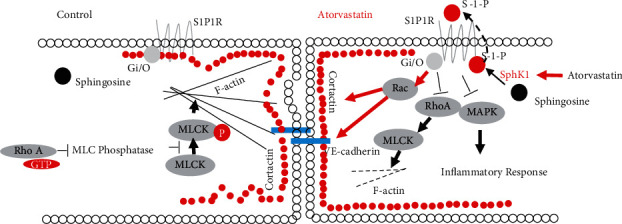
Atorvastatin is a 3-hydroxy-3-methylglutaryl coenzyme A reductase (HMG-CoA reductase) inhibitor and inhibits cholesterol synthesis. Recently, atorvastatin also showed anti-inflammatory effect in acute lung injury, ameliorating pulmonary gas-blood exchanging function. Sphingosine kinase 1 plays a central role in endothelial (EC) cytoskeleton rearrangement and EC barrier integrity regulation. In this study, the role of sphingosine kinase 1 in atorvastatin anti-inflammatory effect against acute lung injury was investigated. Both wild-type (WT) and SphK1-/- mice were challenged with high tidal volume ventilation (40 ml/kg body weight, 65 breathing/min, 4 hours). The acute lung injury was evaluated and the mechanisms were explored. In WT mice, atorvastatin treatment significantly decreased acute lung injury responding to high tidal volume ventilation (HT), including protein, cellular infiltration, and cytokine releasing; comparing to WT mice, SphK1-/- mice showed significantly worsen pulmonary injuries on HT model. Moreover, the atorvastatin-mediated anti-inflammatory effect was diminished in SphK1-/- mice. To further confirm the role of SphK1 in VILI, we then compared the inflammatory response of endothelial cells that were isolated from WT and SphK1-/- mice to cyclic stretching. Similarly, atorvastatin significantly decreased cytokine generation from WT EC responding to cyclic stretching. Atorvastatin also significantly preserved endothelial junction integrity in WT EC against thrombin challenge. However, the inhibitory effect of atorvastatin on cytokine generation induced by cyclic stretching was abolished on SphK1-/- mice EC. The endothelial junction integrity effects of atorvastatin also diminished on SphK1-/- mouse EC. Signal analysis indicated that atorvastatin inhibited JNK activation induced by cyclic stretch. SphK1 knockout also blocked atorvastatin-mediated VE-cadherin junction enhancement. In summary, by inhibition of MAPK activity and maintenance of EC junction homeostasis, SphK1 plays a critical role in atorvastatin-mediated anti-inflammatory effects in both cellular and in vivo model. This study also offers an insight into mechanical stress-mediated acute lung injury and potential therapy in the future.


Conflict of interest statement

The authors declared no potential conflicts of interest with respect to the research, authorship, and/or publication of this article.

Figures

Figure 1
Figure 1
Atorvastatin attenuated VILI-mediated lung permeability but SphK1 knockout reversed this effect. WT and SphK1−/− were treated with atorvastatin overnight as described in Materials and Methods. The next day, the mice underwent high tidal volume ventilation (40 ml/kg, 65 breaths/min, 4 hours). After ventilation, the mouse BAL and lung were harvested for protein, cellular, and histology analysis. (a, b) Compared to spontaneously breathing control mice, HT significantly induced both protein and cells in BAL. Atorvastatin treatment attenuated the HT-mediated induction of protein and cells in BAL. Compared to WT mice, HT-induced SphK1−/− mice had more protein and cell infiltration in the BAL. However, SphK1 knockout significantly abolished the atorvastatin-mediated anti-inflammatory effect in SphK1−/− mice. (c, d) Compared to WT mice, HT-induced SphK1−/− mice had significantly more cell filtration and alveolar structure damage with higher lung injury scores. Moreover, SphK1 knockout reversed atorvastatin-mediated cell filtration inhibitory effect on WT mice (P < 0.05, ∗∗P < 0.01).
Figure 2
Figure 2
Atorvastatin attenuated HT-mediated cytokine generation in BAL, but SphK1 knockout diminished this effect. ELISA was performed on the BAL harvested from both post-HT WT and SphK1−/− mice as described in Materials and Methods. Similar with protein and cell infiltration, HT significantly increased cytokine release in WT mouse BAL. These cytokines included IL6, KC, MIP-1α, MIP-2, and MCP-1, but not IL-1β. HT-mediated cytokine release in WT mouse BAL was significantly attenuated by pretreatment of atorvastatin; HT-mediated cytokine generation was significantly higher in SphK1−/− mouse BAL than that in WT mice. Moreover, SphK1 knockout reversed the anti-inflammatory effect of atorvastatin in cytokine generation (P < 0.05, ∗∗P < 0.01).
Figure 3
Figure 3
The endothelial cell isolation from WT and SphK1−/− mouse lung and identification. The 8-week WT and SphK1−/− mice were sacrificed, and lungs were harvested as described in Materials and Methods. (a) The endothelial cells of WT mouse lung were purified by flow cytometry (CD31+/CD45). (b) The endothelial cells of SphK1−/− mouse lung were isolated and purified (CD31+/CD45). (c) The purified WT and SphK1−/− endothelial cells were cultured on glass slides, and morphological properties were compared, including size, lamellipodia, and junctional properties. (d) qPCR and (e) western blot were performed to determine the sphingosine kinase 1 mRNA and protein level in purified endothelial cells.
Figure 4
Figure 4
Atorvastatin attenuated cyclic stretch- (CS-) mediated cytokine generation from mouse-isolated endothelial cells, but SphK1 knockout reversed this effect. The isolated ECs were seeded on Flexcell plate and cyclic stretched (CS) (18% elongation, 0.5 Hz, 4 hours). After cyclic stretch, the medium was harvested for ELISA. CS significantly increased cytokine release in the WT endothelial medium. These cytokines include IL6, KC, MIP-1α, MIP-2, and MCP-1, but not IL-1β. The CS-mediated cytokine release in WT mouse endothelial medium was significantly attenuated by pretreatment of atorvastatin; CS-mediated cytokine concentrations were significantly higher in SphK1−/− mouse endothelial medium than those in WT mice. Moreover, SphK1 knockout reversed the inhibitory effect of atorvastatin in cytokine generation in endothelial cells. (P < 0.05, ∗∗P < 0.01).
Figure 5
Figure 5
Atorvastatin attenuated cyclic stretch- (CS-) mediated MAPK phosphorylation, and SphK1 knockout reversed this effect. After CS finished at indicated time, the cellular lysates were harvested for western blotting. Compared to the static control, CS significantly induced (b) P38 and (c) JNK phosphorylation in mouse WT ECs. The MAPK phosphorylation was significantly attenuated by pretreatment of atorvastatin. Compared with WT endothelial cells, SphK1−/− cells showed enhanced CS-mediated P38 and JNK phosphorylation. Moreover, CS-mediated P38 and JNK phosphorylation levels in SphK1−/− ECs were significantly higher than those in WT ECs after treatment of atorvastatin. (P < 0.05, ∗∗P < 0.01).
Figure 6
Figure 6
JNK plays a critical role in sphingosine kinase 1-mediated inflammatory response to cyclic stretch. The isolated ECs were seeded on Flexcell plates overnight in the presence of the indicated inhibitors. After cyclic stretching (CS) (18% elongation, 0.5 Hz, 4 hours) as described in Figure 4, the medium was harvested for ELISA. (a) IL6 and KC assay showed that SphK1 knockout significantly enhanced CS-mediated IL6 and KC generation. The SphK1 knockout-mediated IL6 generation enhancement was not significantly abolished by PD98059 but partially by SB203580 and completely by JNK inhibitor II. (b) The SphK1 knockout-mediated KC generation enhancement was not significantly abolished by PD98059 and SB203580, but completely by JNK inhibitor II (P < 0.05, ∗∗P < 0.01).
Figure 7
Figure 7
Atorvastatin did not change SphK1 expression in mouse EC but enhance EC junction integrity. (a) The ECs isolated from WT and SphK1−/− mice were cultured in 6-well plates. The protein levels of SphK1 and SphK2 were determined by western blotting. (b) The ECs isolated from WT or SphK1−/− mice were cultured on glass slides in a 12-well plate. After treatment with 10 μM atorvastatin for 16 hours, the cells were challenged by 1 U/ml thrombin for 1 hour. Immunostaining was performed with VE-cadherin antibody as described in Materials and Methods. (c) Monolayer endothelial cells were cultured on transwell inserts and treated with 10 μM atorvastatin for 16 hours. The next day, the cells were treated with 1 U/ml thrombin for 1 hour in the presence of FITC-dextran. The FITC-dextran fluorescent density in the bottom chamber was measured at 485/538 nm (P < 0.05).
Figure 8
Figure 8
Schematic description of the potential role of SphK1 in atorvastatin-mediated anti-inflammatory effect. Under control condition, sphingosine exists as an inactive form and there is balance between the stress fiber system and the cortactin ring system. When atorvastatin is added, stress fiber is inactivated, the cortical ring is enhanced, and the VE-cadherin junction is tightened. Meanwhile, atorvastatin inhibits the MAPK signal pathway and attenuates inflammatory responses. Sphingosine kinase 1 is a mediator in these processes.

Similar articles

Cited by

References

    1. Toth P. P., Banach M. Statins: then and now. Methodist DeBakey Cardiovascular Journal . 2019;15(1):23–31. doi: 10.14797/mdcj-15-1-23. - DOI - PMC - PubMed
    1. Oesterle A., Laufs U., Liao J. K. Pleiotropic effects of statins on the cardiovascular system. Circulation Research . 2017;120(1):229–243. doi: 10.1161/CIRCRESAHA.116.308537. - DOI - PMC - PubMed
    1. Mager D. R. Statins. Home Healthcare Now . 2016;34(7):388–393. doi: 10.1097/NHH.0000000000000426. - DOI - PubMed
    1. Siempos I. I., Maniatis N. A., Kopterides P., et al. Pretreatment with atorvastatin attenuates lung injury caused by high-stretch mechanical ventilation in an isolated rabbit lung model. Critical Care Medicine . 2010;38(5):1321–1328. doi: 10.1097/CCM.0b013e3181d9dad6. - DOI - PubMed
    1. Tremoulet A. H. The role of statins in inflammatory vasculitides. Autoimmunity . 2015;48(3):177–180. doi: 10.3109/08916934.2015.1027818. - DOI - PubMed

MeSH terms