Review
. 2021 Dec 31;79(1):18.
doi: 10.1007/s00018-021-04075-3.

Meiotic sex chromosome inactivation and the XY body: a phase separation hypothesis

Affiliations
Review

Meiotic sex chromosome inactivation and the XY body: a phase separation hypothesis

Kris G Alavattam et al. Cell Mol Life Sci. .

Abstract

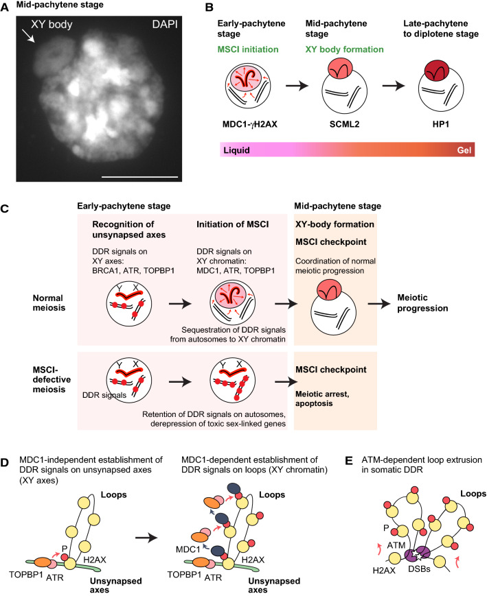
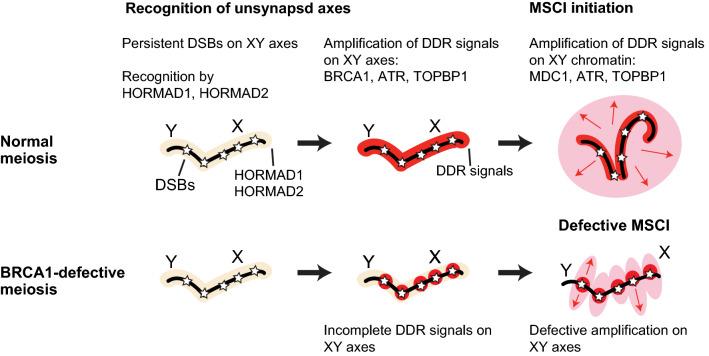
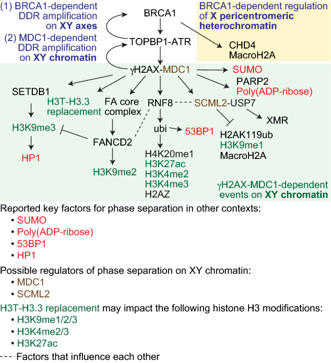
In mammalian male meiosis, the heterologous X and Y chromosomes remain unsynapsed and, as a result, are subject to meiotic sex chromosome inactivation (MSCI). MSCI is required for the successful completion of spermatogenesis. Following the initiation of MSCI, the X and Y chromosomes undergo various epigenetic modifications and are transformed into a nuclear body termed the XY body. Here, we review the mechanisms underlying the initiation of two essential, sequential processes in meiotic prophase I: MSCI and XY-body formation. The initiation of MSCI is directed by the action of DNA damage response (DDR) pathways; downstream of the DDR, unique epigenetic states are established, leading to the formation of the XY body. Accumulating evidence suggests that MSCI and subsequent XY-body formation may be driven by phase separation, a physical process that governs the formation of membraneless organelles and other biomolecular condensates. Thus, here we gather literature-based evidence to explore a phase separation hypothesis for the initiation of MSCI and the formation of the XY body.

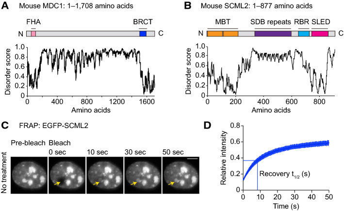
Keywords: Epigenetics; Germ cells; Germline; Liquid–liquid phase separation; Sex body; Sex chromosomes.


Conflict of interest statement

The authors declare no competing interests.

Figures

Fig. 1
Fig. 1
Mechanisms of MSCI initiation. A DAPI counterstaining of a “3D slide,” i.e., a slide prepared such that the gross three-dimensional organization of chromatin is preserved [94]. The dashed circle indicates the XY body. Scale bar: 10 µm. The image is originally from Abe et al. 2020 [30]. B Model of phase separation of the sex chromosomes. Key proteins involved at each step of the process are shown. C Model of the MSCI checkpoint: the physical seclusion of DDR factors from autosomes to the XY body is a critical checkpoint in the progression of meiosis and the development of gametes. At the onset of MSCI, DDR factors (shown as red) are excluded from autosomes and sequestered to the sex chromosomes. The physical seclusion of DDR factors on/at the XY body, which may involve phase separation, is a critical step in the MSCI checkpoint in the mid-pachytene stage of meiotic prophase I. While the MSCI checkpoint ensures meiotic stage progression in normal meiosis, the abolishment of MSCI enables the ectopic retention of DDR signals on/at autosomes; in turn, this triggers complete meiotic arrest and cell death in response to the checkpoint. D Model for the initiation of MSCI. ATR and its activator, TOPBP1, are recruited to unsynapsed axes in an MDC1- and H2AX-Y142-independent manner, resulting in phosphorylation of H2AX (γH2AX) on axes (left). Then, γH2AX recruits MDC1, which facilitates the progressive recruitment of ATR and TOPBP1, resulting in γH2AX and MDC1 spreading throughout loops (right). E Model for ATM-dependent loop extrusion in the somatic DDR. DSBs trigger the recruitment of ATM, which phosphorylates H2AX. As loop extrusion progresses, DNA passes by ATM enzymes at sites of DSBs, facilitating the phosphorylation of histones across large tracts of DNA
Fig. 2
Fig. 2
Depiction of the role of BRCA1 in meiosis. Model for BRCA1-dependent amplification of DDR signals along the XY axes followed by the initiation of MSCI
Fig. 3
Fig. 3
Model for molecular mechanisms in MSCI, including roles for DDR factors, other chromatin-associated proteins, and histone modifications. MSCI is initiated by BRCA1-dependent DDR amplification on the XY axes (1). BRCA1 has an additional function in the establishment of X pericentromeric heterochromatin (yellow box). MDC1-dependent DDR amplification subsequently takes place on XY chromatin (2; green box). Here, we focus on DDR signaling events, recruitment of chromatin factors such as HP1, and histone modifications that occur on XY chromatin. Since the formation of the XY body may involve phase separation, it should be noted that SUMO, Poly(ADP-ribose), 53BP1, and HP1 (shown in red), all of which are related to MSCI, and which are regulated downstream of MDC1, are reported to be key factors for phase separation in other contexts. MDC1 and SCML2 (shown in brown) are possible regulators of phase separation on XY chromatin, as discussed in this review. H3T-H3.3 replacement may impact the following histone H3 modifications: H3K9me1/2/3, H3K4me2/3, and H3K27ac (shown in green). Dashed lines signify factors that influence each other. A key of color codes for various factors is shown at the bottom
Fig. 4
Fig. 4
A phase separation hypothesis for the initiation of MSCI and for XY-body formation. A, B Predictions of protein disorder in MDC1 (A) and SCML2 (B; IUPRED score, Dosztányi et al., 2018 [161]). C Representative live images of FRAP assays in mK4 cells expressing EGFP-SCML2. Bars: 5 μm. The images are originally from Maezawa et al., 2018 [140]. Arrows show the site of photobleaching. D Relative intensity from FRAP assays of full-length SCML2. Recovery represents 50% of the plateaued intensity: t1/2 (s). Error bars for FRAP curves and recovery t1/2 represent 95% confidence intervals of the mean. The data are originally from Maezawa et al. 2018 [140]

References

    1. Boeynaems S, Alberti S, Fawzi NL, Mittag T, Polymenidou M, Rousseau F, Schymkowitz J, Shorter J, Wolozin B, Van Den Bosch L, Tompa P, Fuxreiter M. Protein phase separation: a new phase in cell biology. Trends Cell Biol. 2018;28(6):420–435. doi: 10.1016/j.tcb.2018.02.004. - DOI - PMC - PubMed
    1. Zhao YG, Zhang H. Phase separation in membrane biology: the interplay between membrane-bound organelles and membraneless condensates. Dev Cell. 2020;55(1):30–44. doi: 10.1016/j.devcel.2020.06.033. - DOI - PubMed
    1. Schmidt HB, Görlich D. Transport selectivity of nuclear pores, phase separation, and membraneless organelles. Trends Biochem Sci. 2016;41(1):46–61. doi: 10.1016/j.tibs.2015.11.001. - DOI - PubMed
    1. Brangwynne CP, Mitchison TJ, Hyman AA. Active liquid-like behavior of nucleoli determines their size and shape in Xenopus laevis oocytes. Proc Natl Acad Sci USA. 2011;108(11):4334–4339. doi: 10.1073/pnas.1017150108. - DOI - PMC - PubMed
    1. Strom AR, Emelyanov AV, Mir M, Fyodorov DV, Darzacq X, Karpen GH. Phase separation drives heterochromatin domain formation. Nature. 2017;547(7662):241–245. doi: 10.1038/nature22989. - DOI - PMC - PubMed