. 2022 Mar;113(3):864-874.
doi: 10.1111/cas.15259. Epub 2022 Jan 17.

Cancer immunotherapy using artificial adjuvant vector cells to deliver NY-ESO-1 antigen to dendritic cells in situ

Affiliations

Cancer immunotherapy using artificial adjuvant vector cells to deliver NY-ESO-1 antigen to dendritic cells in situ

Shin-Ichiro Fujii et al. Cancer Sci. 2022 Mar.

Abstract

NY-ESO-1 is a cancer/testis antigen expressed in various cancer types. However, the induction of NY-ESO-1-specific CTLs through vaccines is somewhat difficult. Thus, we developed a new type of artificial adjuvant vector cell (aAVC-NY-ESO-1) expressing a CD1d-NKT cell ligand complex and a tumor-associated antigen, NY-ESO-1. First, we determined the activation of invariant natural killer T (iNKT) and natural killer (NK) cell responses by aAVC-NY-ESO-1. We then showed that the NY-ESO-1-specific CTL response was successfully elicited through aAVC-NY-ESO-1 therapy. After injection of aAVC-NY-ESO-1, we found that dendritic cells (DCs) in situ expressed high levels of costimulatory molecules and produced interleukn-12 (IL-12), indicating that DCs undergo maturation in vivo. Furthermore, the NY-ESO-1 antigen from aAVC-NY-ESO-1 was delivered to the DCs in vivo, and it was presented on MHC class I molecules. The cross-presentation of the NY-ESO-1 antigen was absent in conventional DC-deficient mice, suggesting a host DC-mediated CTL response. Thus, this strategy helps generate sufficient CD8+ NY-ESO-1-specific CTLs along with iNKT and NK cell activation, resulting in a strong antitumor effect. Furthermore, we established a human DC-transferred NOD/Shi-scid/IL-2γcnull immunodeficient mouse model and showed that the NY-ESO-1 antigen from aAVC-NY-ESO-1 was cross-presented to antigen-specific CTLs through human DCs. Taken together, these data suggest that aAVC-NY-ESO-1 has potential for harnessing innate and adaptive immunity against NY-ESO-1-expressing malignancies.

Keywords: NKT cell; cancer; cytotoxic T cell; dendritic cell; immunotherapy.


Figures

FIGURE 1
FIGURE 1
Activation of innate immunity by NY‐ESO‐1‐expressing artificial adjuvant cells (aAVC‐NY‐ESO‐1). A, aAVC‐NY‐ESO‐1 cells were established by loading NIH3T3 vector cells with α‐galactosylceramide (α‐GalCer) and coelectroporation with NY‐ESO‐1 and murine CD1d mRNA. B‐D, NY‐ESO‐1 protein levels and CD1d surface expression were assessed using western blotting (B) and flow cytometry (C), respectively (red, aAVC‐ NY‐ESO‐1; bold, NIH3T3; shaded, isotype). D, α‐GalCer presentation by aAVC‐NY‐ESO‐1. aAVC‐NY‐ESO‐1 were cocultured with the Vα14 invariant natural killer T (iNKT) cell hybridoma 1.2 for 24 h, and interleukin‐2 (IL‐2) production in the culture supernatant was evaluated by ELISA (n = 4; mean ± SEM) ***P < .001 (Mann‐Whitney). E, iNKT and natural killer (NK) cell activation. Mice were injected with 5 × 105 aAVC‐NY‐ESO‐1 i.v. Spleens were removed 6 h later, and iNKT as well as NK cells were analyzed by staining with CD19‐antigen presenting cells (APC) and CD1d‐dimer+ phycoerythrin (PE) or CD3‐FITC and NK1.1‐APC. These cells were simultaneously stained with either surface CD69‐PE or intracellular interferon‐γ‐PE and evaluated through flow cytometry (n = 4). F, Antitumor immunity by innate lymphocytes. Mice were injected with 3 × 105 B16 melanoma cells i.v. and, 3 h later, were inoculated with 5 × 105 aAVC‐NY‐ESO‐1. The number of lung metastases was determined 14 d later (n = 5 per group, mean ± SEM). In some experiments, NK cells were depleted using anti‐asialo GM1 Ab. ***P < .001 (Tukey’s test) (aAVC‐immunized vs others)
FIGURE 2
FIGURE 2
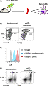
NY‐ESO‐1‐expressing artificial adjuvant cells (aAVC‐NY‐ESO‐1) lead to the functional maturation of dendritic cells (DCs) in situ. A, Three million carboxyfluorescein succinimidyl ester (CFSE)‐labeled aAVC‐NY‐ESO‐1 were injected i.v. into C57BL/6 mice. Twelve hours later, splenic DCs were analyzed. B, Uptake of CFSE+ aAVCs by murine CD11c+ splenic DCs was measured based on CFSE+ gating on CD11c+ class II+ by flow cytometry. C, DCs were analyzed for the upregulation of CD86 expression after gating on CD11c+ at 12 h using CD11c‐antigen presenting cells and CD86‐phycoerythrin (isotype control, shaded; nonimmunized mice, blue; aAVC‐immunized mice, red). Data are representative of three independent experiments (n = 3 per group). D, Interleukin‐12 (IL‐12) production by conventional DCs in vivo was evaluated through intracellular staining (n = 3 per group, mean ± SEM)
FIGURE 3
FIGURE 3
NY‐ESO‐1‐specific CD8+ T cell response induced by NY‐ESO‐1‐expressing artificial adjuvant cells (aAVC‐NY‐ESO‐1). A, Mice were immunized with 5 × 105 aAVC‐NY‐ESO‐1 i.v. One week later, NY‐ESO‐1‐specific CD8+ T cells were analyzed by interferon‐γ (IFN‐γ) enzyme‐linked immune absorbent spot (ELISPOT) assay. CD8+ T cells were obtained from the spleen and cultured with splenic CD11c+ dendritic cells (DCs) from naïve mice in the presence or absence of the NY‐ESO‐1 peptide pool for 24 h. B, NY‐ESO‐1‐specific CD8+ T cell response in aAVC‐NY‐ESO‐1‐immunized mice and nonimmunized mice (n = 5 per group, mean ± SEM). ***P < .001 (aAVC‐NY‐ESO‐1 CD8T+CD11c+peptivator vs others, Tukey’s test). C, NY‐ESO‐1‐specific CD8+ T cell response in conventional DC (cDC)‐depleted mice. To test the role of cDCs in the generation of NY‐ESO‐1‐specific CD8+ T cells, WT bone marrow (BM) or zDC‐DTR BM chimera mice were treated with diphtheria toxin (DT) and then immunized with aAVC‐NY‐ESO‐1 (n = 4 per group, mean ± SEM). *P < .05 (WT BM chimera vs zDC‐DTR BM chimera, Mann‐Whitney). ns, not significant; SFC, spot forming cell
FIGURE 4
FIGURE 4
Antitumor response induced by NY‐ESO‐1‐expressing artificial adjuvant cells (aAVC‐NY‐ESO‐1). Antitumor response was evaluated in a prophylactic and a therapeutic vaccination model. A, Mice were immunized with 5 × 105 aAVC‐OVA i.v. or OVA peptide plus complete Freund’s adjuvant (CFA) and challenged with 1 × 105 MO4 s.c. at day 14 (d14). (n = 5‐6 per group, mean ± SEM). *P < .001. ns, not significant (Mann‐Whitney). MO4, black; CFA/OVApep, blue; aAVC‐OVA, red. B, Mice were inoculated with 5 × 105 MO4 s.c. and treated with 5 × 105 aAVC‐OVA or OVA peptide plus CFA on d10. Tumor size was measured at the indicated time points (n = 5‐6 per group, mean ± SEM). **P < .01 (Mann‐Whitney). MO4, black; CFA/OVApep, blue; aAVC‐OVA, red. C, D, Mice were immunized with 5 × 105 aAVC‐NY‐ESO‐1 i.v. and challenged with 1 × 105 NY‐ESO‐1‐expressing B16 (B16‐NY‐ESO‐1) (C, left) or B16 (C, right) s.c. at d14 (n = 7‐8 per group, mean ± SEM). *P < .001. ns, not significant (Mann‐Whitney). B16 or B16‐NY‐ESO‐1, black; aAVC‐NY‐ESO‐1, red. D, Mice were inoculated with 2 × 105 B16‐NY‐ESO‐1 s.c. and treated with 5 × 105 aAVC‐NY‐ESO‐1 on d10. Tumor size was measured at the indicated time points (n = 6‐7 per group, mean ± SEM). **P < .01 (Mann‐Whitney). B16 or B16‐NY‐ESO‐1, black; aAVC‐NY‐ESO‐1, red
FIGURE 5
FIGURE 5
Human T cell activation by dendritic cells in NOG mice immunized with human NY‐ESO‐1‐expressing artificial adjuvant cells (aAVC‐NY‐ESO‐1). A, Human aAVC‐NY‐ESO‐1 were established by loading HEK293sf cells with α‐galactosylceramide (α‐GalCer) and coelectroporation with NY‐ESO‐1 as well as human CD1d mRNA. Recombinant NY‐ESO‐1 protein expression in aAVC‐NY‐ESO‐1 (left) and hCD1d expression (right). B, α‐GalCer presentation by human aAVC‐NY‐ESO‐1. Human natural killer T (NKT) cells were cocultured with HEK293sf or aAVC‐NY‐ESO‐1 for 24 h. Interferon‐γ (IFN‐γ) production was analyzed by ELISA (n = 4 per group, mean ± SEM). *P < .05 (Mann‐Whitney). C, Activation of NKT cells by human type aAVC‐NY‐ESO‐1 in C57BL/6 mice. Mice were immunized with 5 × 105 aAVC‐NY‐ESO‐1 i.v. Three days later, the frequency of invariant NKT (iNKT) cells in the spleen was analyzed by flow cytometry. D, CD8+ T cell activation in humanized mice. NY‐ESO‐1 TCR‐T cells, NKT cells, and dendritic cells (DCs) were prepared from the same HLA‐A2+ healthy donor. The treatment protocol is shown. These T cells (NY‐ESO‐1 TCR‐T) were labeled with carboxyfluorescein succinimidyl ester (CFSE) and injected into NOG mice. Three hours later, monocyte‐derived immature DCs (imDC) and iNKT cells were transferred, together with or without aAVC‐NY‐ESO‐1. Five days later, the proliferation of NY‐ESO‐1 TCR‐T cells was evaluated by flow cytometry. Histograms from one representative donor are shown (n = 3)

References

    1. Coulie PG, Van den Eynde BJ, van der Bruggen P, Boon T. Tumour antigens recognized by T lymphocytes: at the core of cancer immunotherapy. Nat Rev Cancer. 2014;14:135‐146. - PubMed
    1. Schumacher TN, Schreiber RD. Neoantigens in cancer immunotherapy. Science. 2015;348:69‐74. - PubMed
    1. Anderson KG, Stromnes IM, Greenberg PD. Obstacles posed by the tumor microenvironment to T cell activity: a case for synergistic therapies. Cancer Cell. 2017;31:311‐325. - PMC - PubMed
    1. Wherry EJ, Kurachi M. Molecular and cellular insights into T cell exhaustion. Nat Rev Immunol. 2015;15:486‐499. - PMC - PubMed
    1. Chen YT, Scanlan MJ, Sahin U, et al. A testicular antigen aberrantly expressed in human cancers detected by autologous antibody screening. Proc Natl Acad Sci USA. 1997;94:1914‐1918. - PMC - PubMed

MeSH terms