

Canine endotheliitis: Clinical characteristics, advanced imaging features, and treatment
- PMID: 34971485
- PMCID: PMC9243184
- DOI: 10.1111/vop.12967
Canine endotheliitis: Clinical characteristics, advanced imaging features, and treatment
Abstract
Objective: To describe the clinical findings, multimodal corneal imaging features and treatment in canine patients diagnosed with endotheliitis.
Animals studied: Four canine patients met inclusion criteria for bilateral corneal disease with endothelial inflammation and secondary corneal edema that responded to topical anti-inflammatory treatment.
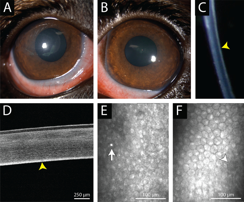
Methods: The patients selected underwent a complete ophthalmic examination with emphasis on the cornea including ultrasound pachymetry (USP), Fourier-domain optical coherence tomography (FD-OCT), in vivo confocal microscopy (IVCM), and digital slit lamp photography.
Results: All patients in this study demonstrated thickened corneas due to edema with USP and FD-OCT. With IVCM, mild to severe polymegathism and pleomorphism of corneal endothelial cells, reduced endothelial cell density, hyperreflective keratic precipitates (KPs), and extracellular debris as well as hyporeflective pseudoguttata were observed. With FD-OCT, hyperreflective KPs were commonly observed on the inferior cornea. Clinical examination and advanced imaging results were consistent with a diagnosis of endotheliitis. All patients initially responded to topical anti-inflammatory treatment and required continued therapy; two patients also received topical netarsudil, a rho-associated coiled-coil kinase inhibitor.
Conclusion: Endotheliitis should be considered for canine patients with bilateral edema that is most severe in the inferior cornea. Careful inspection of Descemet's membrane-endothelial complex should be performed for KPs or inflammatory debris. Chronic administration of topical anti-inflammatories may be necessary to prevent flare-ups of endotheliitis.
Keywords: ROCK inhibitor; corneal edema; endotheliitis; in vivo confocal microscopy; optical coherence tomography.
© 2021 American College of Veterinary Ophthalmologists.
Conflict of interest statement
CONFLICT OF INTEREST
While the netarsudil (Rhopressa®) was donated by Aerie Pharmaceuticals and could be viewed as a possible conflict of interest, we highlight that they had no input in the design, analysis or interpretation of the data presented in the manuscript or the manuscript itself.
Figures


Similar articles
-
Superficial Keratectomy and Conjunctival Advancement Hood Flap (SKCAHF) for the Management of Bullous Keratopathy: Validation in Dogs With Spontaneous Disease.Cornea. 2016 Oct;35(10):1295-304. doi: 10.1097/ICO.0000000000000966. Cornea. 2016. PMID: 27538190 Free PMC article.
-
Phenotypic Characterization of Corneal Endothelial Dystrophy in German Shorthaired and Wirehaired Pointers Using In Vivo Advanced Corneal Imaging and Histopathology.Cornea. 2018 Jan;37(1):88-94. doi: 10.1097/ICO.0000000000001431. Cornea. 2018. PMID: 29077583 Free PMC article.
-
In Vivo Imaging of Corneal Endothelial Dystrophy in Boston Terriers: A Spontaneous, Canine Model for Fuchs' Endothelial Corneal Dystrophy.Invest Ophthalmol Vis Sci. 2016 Jul 1;57(9):OCT495-503. doi: 10.1167/iovs.15-18885. Invest Ophthalmol Vis Sci. 2016. PMID: 27454658 Free PMC article.
-
The ROCK inhibitor netarsudil in the treatment of corneal endothelial decompensation caused by corneal endotheliitis: A case report and literature review.Int Immunopharmacol. 2024 Jul 30;136:112195. doi: 10.1016/j.intimp.2024.112195. Epub 2024 May 30. Int Immunopharmacol. 2024. PMID: 38820965 Review.
-
A Review of Corneal Endotheliitis and Endotheliopathy: Differential Diagnosis, Evaluation, and Treatment.Ophthalmol Ther. 2019 Jun;8(2):195-213. doi: 10.1007/s40123-019-0169-7. Epub 2019 Mar 11. Ophthalmol Ther. 2019. PMID: 30859513 Free PMC article. Review.
Cited by
-
Topical netarsudil for the treatment of primary corneal endothelial degeneration in dogs.Sci Rep. 2024 Mar 14;14(1):6238. doi: 10.1038/s41598-024-56084-4. Sci Rep. 2024. PMID: 38485975 Free PMC article.
References
-
- Thomasy SM, Cortes DE, Hoehn AL, Calderon AC, Li JY, Murphy CJ. In Vivo Imaging of Corneal Endothelial Dystrophy in Boston Terriers: A Spontaneous, Canine Model for Fuchs’ Endothelial Corneal Dystrophy. Investigative ophthalmology & visual science. 2016;57(9):OCT495–503. doi:10.1167/iovs.15-18885 - DOI - PMC - PubMed
-
- Dubielzig RR, Ketring K, McLellan GJ, Albert DM. Chapter 8: Diseases of the cornea and sclera. In: Dubielzig RR, Ketring K, McLellan GJ, Albert DM, eds. Veterinary Ocular Pathology. W.B. Saunders; 2010:201–243. doi:10.1016/B978-0-7020-2797-0.00008-4 - DOI
-
- Gwin RM, Lerner I, Warren JK, Gum G. Decrease in canine corneal endothelial cell density and increase in corneal thickness as functions of age. Invest Ophthalmol Vis Sci. 1982;22(2):267–271. - PubMed
Publication types
MeSH terms
Grants and funding
LinkOut - more resources
Full Text Sources
Medical
Miscellaneous
