. 2022 Feb;96(2):613-624.
doi: 10.1007/s00204-021-03197-8. Epub 2022 Jan 1.

Pharmacological inhibition of MERTK induces in vivo retinal degeneration: a multimodal imaging ocular safety assessment

Affiliations

Pharmacological inhibition of MERTK induces in vivo retinal degeneration: a multimodal imaging ocular safety assessment

Gregory Hamm et al. Arch Toxicol. 2022 Feb.

Abstract

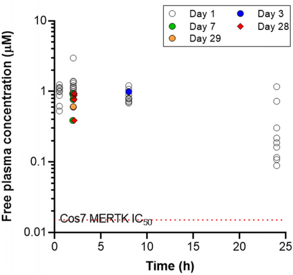
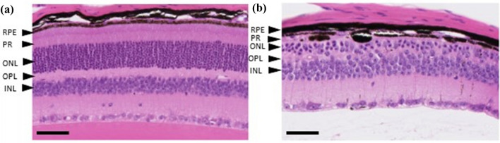
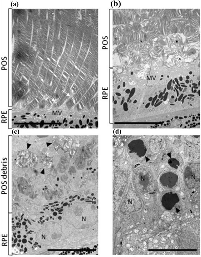
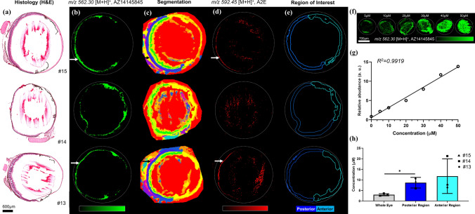
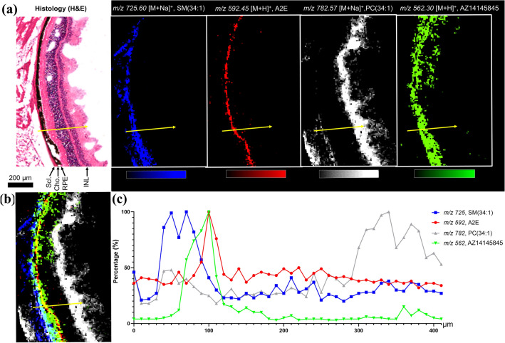
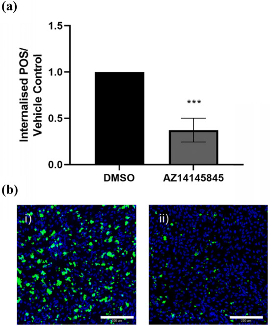
The receptor tyrosine kinase, MERTK, plays an essential role in homeostasis of the retina via efferocytosis of shed outer nuclear segments of photoreceptors. The Royal College of Surgeons rat model of retinal degeneration has been linked to loss-of-function of MERTK, and together with the MERTK knock-out mouse, phenocopy retinitis pigmentosa in humans with MERTK mutations. Given recent efforts and interest in MERTK as a potential immuno-oncology target, development of a strategy to assess ocular safety at an early pre-clinical stage is critical. We have applied a state-of-the-art, multi-modal imaging platform to assess the in vivo effects of pharmacological inhibition of MERTK in mice. This involved the application of mass spectrometry imaging (MSI) to characterize the ocular spatial distribution of our highly selective MERTK inhibitor; AZ14145845, together with histopathology and transmission electron microscopy to characterize pathological and ultra-structural change in response to MERTK inhibition. In addition, we assessed the utility of a human retinal in vitro cell model to identify perturbation of phagocytosis post MERTK inhibition. We identified high localized total compound concentrations in the retinal pigment epithelium (RPE) and retinal lesions following 28 days of treatment with AZ14145845. These lesions were present in 4 of 8 treated animals, and were characterized by a thinning of the outer nuclear layer, loss of photoreceptors (PR) and accumulation of photoreceptor outer segments at the interface of the RPE and PRs. Furthermore, the lesions were very similar to that shown in the RCS rat and MERTK knock-out mouse, suggesting a MERTK-induced mechanism of PR cell death. This was further supported by the observation of reduced phagocytosis in the human retinal cell model following treatment with AZ14145845. Our study provides a viable, translational strategy to investigate the pre-clinical toxicity of MERTK inhibitors but is equally transferrable to novel chemotypes.

Keywords: MERTK; Mass spectrometry imaging; Ocular electron microscopy; Ocular histopathology; Retinal degeneration; TAM kinases.


Figures

Fig. 1
Fig. 1
Free plasma concentration of AZ14145845 on day 1, 3, 7, 28 and 29 following 200 mg/kg BID dosing to C57BL/6 J mice
Fig. 2
Fig. 2
Retinal histopathology from FFPE H&E sections. a Animal no. 5 given vehicle only showing the normal layered structure of the retina, including Retinal Pigment Epithelium (RPE), Photoreceptors (PR), Outer Nuclear Layer (ONL), Outer Plexiform Layer (OPL) and Inner Nuclear Layer (INL). b Animal no. 10 given AZ14145845 shows marked thinning of the outer retina with loss of nuclei from the ONL and severe thinning of the ONL and PR layers and thinning of the PR containing displaced melanin pigment. Scale Bar = 50 µM
Fig. 3
Fig. 3
TEM micrographs: Ultrastructural changes in retinal layers by Electron Microscopy. a Transmission electron micrograph of interface between POS and RPE cells showing microvilli (MV) or RPE cells interdigitating (white arrows) with well-organised tips of POS in a vehicle-dosed animal. b Interface of RPE microvilli (MV) and POS in a AZ14145845-treated animal showing disorganization of POS and free strands of discs material (*). c Apical surface of RPE cells (N = nuclei) showing normally well-organised POS tips replaced by a zone of debris containing membrane-bound whorls (arrows) or POS material admixed with free degenerating POS material and microvilli. d Nuclei (N) of outer nuclear layer in a AZ14145845-treated animal showing several nuclei (arrows) with condensed chromatin. Scale bar = 10 µm
Fig. 4
Fig. 4
MALDI Mass Spectrometry Imaging analysis of whole eye sections treated with AZ14145845 obtained in positive ion mode at 20 µm spatial resolution. a Histological staining of adjacent eye sections using Haematoxylin and Eosin (H&E). b AZ14145845 molecular images from MSI. The white arrow shows the individual retinal layers. c Spatial segmentation. d A2E molecular images. e Region of interest describing the posterior and the anterior part of the eye used for the quantification of AZ14145845. f Molecular images of AZ14145845 dilution series spotted onto control eye section from 5 to 50 μM. g Calibration curve generated from AZ14145845 relative abundances on the dilution series molecular images. h Quantification of AZ14145845 in the whole eye, posterior and anterior segments in µm. Scale bar = 600 µm
Fig. 5
Fig. 5
MALDI Mass Spectrometry Imaging of retinal layers of an AZ14145845-treated eye section (animal no. 13) at 10 µm spatial resolution showing co-localization of the MERTKi with the molecular marker of the RPE; A2E. a Histology of the eye with corresponding molecular images of Sphingomyelin, SM(34:1) at m/ 725, N-retinylidene-N-retinylethanolamine (A2E) at m/z 592, Phosphatidylcholine PC(34:1) at m/z 782 and MERTKi at m/z 562. b Overlay of molecular images and c line scan data from the region shown by the yellow arrow in (a)
Fig. 6
Fig. 6
FITC-labeled photoreceptor outer segment phagocytosis in human retinal ARPE19 cells. a Quantification of total number of POS as determined by MetaExpress custom module for counting internalised POS particles expressed as fold change over vehicle control (mean ± standard deviation). Statistical significance was calculated using a one-way ANOVA (Graphpad Prism, ***p < 0.0001). b Representative images of (i) vehicle control cells and (ii) AZ14145845-treated cells. Nuclei are stained in blue and FITC-labeled POS are colored green. Scale bar = 200 µm

Comment in

  • Progress in retinal toxicity research.
    Bolt HM, Hengstler JG. Bolt HM, et al. Arch Toxicol. 2022 Feb;96(2):387-388. doi: 10.1007/s00204-022-03222-4. Epub 2022 Jan 31. Arch Toxicol. 2022. PMID: 35098322 Free PMC article. No abstract available.

References

    1. Alexandrov T, Becker M, Deininger SO, et al. Spatial segmentation of imaging mass spectrometry data with edge-preserving image denoising and clustering. J Proteome Res. 2010;9(12):6535–6546. doi: 10.1021/pr100734z. - DOI - PubMed
    1. Anderson DG, Ablonczy Z, Koutalos Y, et al. High resolution MALDI imaging mass spectrometry of retinal tissue lipids. J Am Soc Mass Spectrom. 2014;25:1394–1403. doi: 10.1007/s13361-014-0883-2. - DOI - PMC - PubMed
    1. Boada-Romero E, Martinez J, Heckmann BL, Green DR. The clearance of dead cells by efferocytosis. Nat Rev Mol Cell Biol. 2020;21(7):398–414. doi: 10.1038/s41580-020-0232-1. - DOI - PMC - PubMed
    1. Boughton BA, Thomas ORB, Demarais NJ, Trede D, Swearer SE, Grey AC. Detection of small molecule concentration gradients in ocular tissues and humours. J Mass Spectrom. 2020;55(4):e4460. doi: 10.1002/jms.4460. - DOI - PubMed
    1. Brignole-Baudouin F, Desbenoit N, Hamm G, et al. A new safety concern for glaucoma treatment demonstrated by mass spectrometry imaging of benzalkonium chloride distribution in the eye, an experimental study in rabbits. PLoS ONE. 2012;7(11):e50180. doi: 10.1371/journal.pone.0050180. - DOI - PMC - PubMed

MeSH terms