. 2022 Feb:16:101329.
doi: 10.1016/j.tranon.2021.101329. Epub 2021 Dec 29.

A novel PAK4 inhibitor suppresses pancreatic cancer growth and enhances the inhibitory effect of gemcitabine

Affiliations

A novel PAK4 inhibitor suppresses pancreatic cancer growth and enhances the inhibitory effect of gemcitabine

Hong He et al. Transl Oncol. 2022 Feb.

Abstract

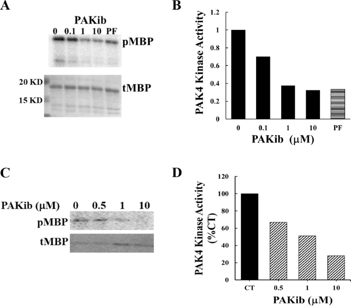
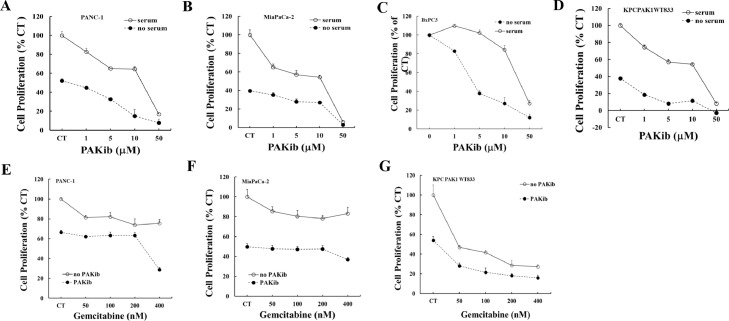
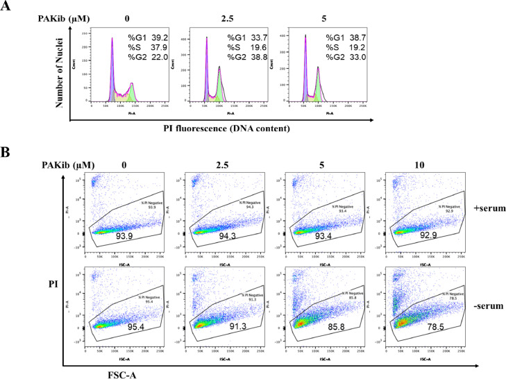
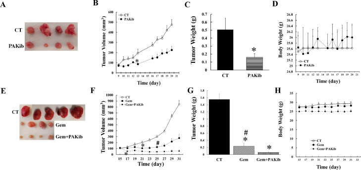
Over 95% of Pancreatic ductal adenocarcinomas (PDA) carry mutations in the oncogene KRas which has been proven to be a difficult drug target. P21-activated kinase 4 (PAK4), acts downstream of KRas, and is overexpressed in PDA contributing to its growth and chemoresistance, and thus becomes an attractive therapeutic target. We have developed a new PAK4 inhibitor, PAKib and tested its effect on pancreatic cancer (PC) cell growth in vitro and in a syngeneic mouse model of PC. PAKib suppressed PC cell growth by inducing cell death and cycle arrest. PAKib inhibited PC growth and enhanced the inhibition by gemcitabine of PC in cell culture and in PC mouse model. PAKib acted through multiple signaling pathways involved in cell cycle checkpoints, apoptosis, cell junction, and focal adhesion. These proof-of-concept studies demonstrated the anti-cancer effect of PAKib alone and in combination with gemcitabine and warrant a further clinical investigation.

Keywords: Gemcitabine; KRas; PAK4; Pancreatic cancer.


Conflict of interest statement

MN and JZ are on the Board of Pakinax Pty Ltd that was involved in the manufacture or the PAKib. JZ is current CEO of Pakinax Pty Ltd. HH is a medical advisor to Pakinax Pty Ltd and was involved in the manufacture of PAKib.

Figures

Fig 1
Fig. 1
PAKib inhibited the kinase activity of PAK4. In an in vitro kinase assay using myelin basic protein (MBP) as substrate, PAKib suppressed the kinase activity of PAK4 (immune-precipitated from PANC-1 cell) with an IC50 value about 500 nM (A, B), and of a recombinant PAK4 (C, D). pMBP: phosphorylated MBP; tMBP: total MBP; CT: control. The density of pMBP from 0 (PAKib, B) or CT (D) were taken as 1 or 100% respectively.
Fig 2
Fig. 2
PAKib decreased PC cell proliferation and enhanced the inhibitory effect of gemcitabine. Human PANC-1 (A), MiaPaCa-2 (B) and BxPC3 (C), and murine KPCPAK1WT833 (D) PC cell lines were treated with PAKib with concentrations indicated in the figures (A-D) with or without serum for 24h. The cell proliferation was determined by MTT assay as described in the Materials and Methods. PAKib dose-dependently decreased the proliferation of both human and murine PC cells. In the combination of PAKib and gemcitabine treatment, PANC-1 (E), MiaPaCa-2 (F) and KPCPAK1WT833 (G) were pre-treated with PAKib (10 μM) for 24h and followed by another 24h treatment of gemcitabine (Gem) with concentrations indicated in the figures (E-G). The date was summarized from three independent experiments. CT: control. The values from non-treated control were taken as 100%.
Fig 3
Fig. 3
PAKib suppressed PC cell growth by inducing cell cycle arrest and cell death. PANC-1 (A) and KPCPAK1WT833 (B) cells were treated with PAKib for 24h before being harvested for propidium iodide (PI) staining and flow cytometry as described in the Materials and Methods. The acquired data were analysed with the FlowJo software. The results were selected and represented from three independent experiments.
Fig 4
Fig. 4
PAKib suppressed PC growth in vivo and enhanced the inhibitory effect of gemcitabine on PC. KPCPAK1WT833 cells (50,000 cell/100 μl DMEM plus 5% FBS) were subcutaneously injected into the back of the right hind leg of a mouse. The mice were treated with PAKib (40 mg/Kg) by a subcutaneous injection (s.i.) every second day when tumour volume reached above 70 mm3 in set one (A-D) experiment where the initial average tumour volumes were 77.51±12.27 mm3 and 70.77±11.41 mm3 for control (CT, n=4) and PAKib (n=4) respectively (B). In set two experiment (E-H), when tumour volume reached 100 mm3, mice were treated with gemcitabine (Gem, 25 mg/Kg) alone every fourth day by intraperitoneal injection (i.p.) or Gem plus PAKic (40 mg/Kg, s.i. every second day). For set two experiment, the initial tumour volumes were 106.34+12.48 mm3, 113.12+13.82 mm3, and 114.53+3.94 mm3 for CT (n=5), Gem alone (n=4) and Gem plus PAKib (n=4) respectively (F). *, p<0.05 compared to CT; #, p<0.05 compared to Gem plus PAKib.
Fig 5
Fig. 5
PAKib induced changes in phosphosites and the phosphorylated proteins. PANC-1 cells were treated with PAKib (10 μM) for 48h before the cells were lysed and the resultant cell lysates were subjected to a proteomic analysis as described in the Material and Methods. 4 repeated samples for control (CT) and PAKib-treated separately were applied in the experiment. The raw data generated through a Mass spectrum were processed using MaxQuant software, and resultant data were input into a Perseus software (version 1.6.14.0) where the changes in phosphosites and related phosphorylated proteins were identified. Statistically significant phosphosites were calculated and the mapped proteins were shown in the Volcano plot (A). The phosphosites with significant changes were used to generate a heatmap (B). Green represented the phosphorylated proteins decreased by PAKib compared to CT while Red PAKib-increased phosphorylated proteins. T: PAKib-treated; C: control
Fig 6
Fig. 6
PAKib affected intracellular pathways. The intracellular pathways affected by PAKib were calculated and identified using Cytoscape software. The data with significant changes in phosphosites generated through Perseus was formatted and entered to Cytoscape software where the related network pathways (solid grey lines) were identified, and the extended network pathways (red dotted lines) were also deduced. The red notes represented PAKib-increased phosphosites in related proteins while the green notes the PAKib-decreased phosphosites in related proteins.

References

    1. Siegel R.L., Miller K.D., Jemal A. Cancer statistics, 2019. CA Cancer J. Clin. 2019;69:7–34. - PubMed
    1. Biankin AV, Waddell N, Kassahn KS, Gingras MC, Muthuswamy LB, Johns AL, Miller DK, Wilson PJ, Patch AM, Wu J, Chang DK, Cowley MJ, Gardiner BB, Song S, Harliwong I, Idrisoglu S, Nourse C, Nourbakhsh E, Manning S, Wani S, Gongora M, Pajic M, Scarlett CJ, Gill AJ, Pinho AV, Rooman I, Anderson M, Holmes O, Leonard C, Taylor D, Wood S, Xu Q, Nones K, Fink JL, Christ A, Bruxner T, Cloonan N, Kolle G, Newell F, Pinese M, Mead RS, Humphris JL, Kaplan W, Jones MD, Colvin EK, Nagrial AM, Humphrey ES, Chou A, Chin VT, Chantrill LA, Mawson A, Samra JS, Kench JG, Lovell JA, Daly RJ, Merrett ND, Toon C, Epari K, Nguyen NQ, Barbour A, Zeps N, I Australian Pancreatic Cancer Genome. Kakkar N, Zhao F, Wu YQ, Wang M, Muzny DM, Fisher WE, Brunicardi FC, Hodges SE, Reid JG, Drummond J, Chang K, Han Y, Lewis LR, Dinh H, Buhay CJ, Beck T, Timms L, Sam M, Begley K, Brown A, Pai D, Panchal A, Buchner N, De Borja R, Denroche RE, Yung CK, Serra S, Onetto N, Mukhopadhyay D, Tsao MS, Shaw PA, Petersen GM, Gallinger S, Hruban RH, Maitra A, Iacobuzio-Donahue CA, Schulick RD, Wolfgang CL, Morgan RA, Lawlor RT, Capelli P, Corbo V, Scardoni M, Tortora G, Tempero MA, Mann KM, Jenkins NA, Perez-Mancera PA, Adams DJ, Largaespada DA, Wessels LF, Rust AG, Stein LD, Tuveson DA, Copeland NG, Musgrove EA, Scarpa A, Eshleman JR, Hudson TJ, Sutherland RL, Wheeler DA, Pearson JV, McPherson JD, Gibbs RA, Grimmond SM. Pancreatic cancer genomes reveal aberrations in axon guidance pathway genes. Nature. 2012;491:399–405. - PMC - PubMed
    1. Bournet B., Buscail C., Muscari F., Cordelier P., Buscail L. Targeting KRAS for diagnosis, prognosis, and treatment of pancreatic cancer: Hopes and realities. Eur. J. Cancer. 2016;54:75–83. - PubMed
    1. Wang K., Baldwin G.S., Nikfarjam M., He H. p21-activated kinase signalling in pancreatic cancer: New insights into tumour biology and immune modulation. World J. Gastroenterol. 2018;24:3709–3723. - PMC - PubMed
    1. Yeo D., He H., Baldwin G.S., Nikfarjam M. The role of p21-activated kinases in pancreatic cancer. Pancreas. 2015;44:363–369. - PubMed