. 2022 Feb 1;121(3):347-360.
doi: 10.1016/j.bpj.2021.12.043. Epub 2021 Dec 30.

The Bak core dimer focuses triacylglycerides in the membrane

Affiliations

The Bak core dimer focuses triacylglycerides in the membrane

Nicholas A Smith et al. Biophys J. .

Abstract

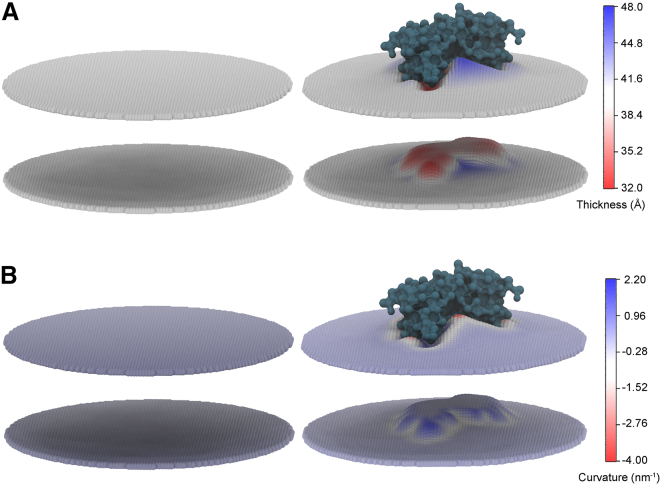
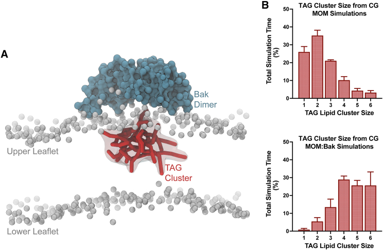
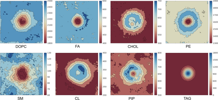
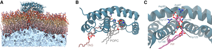
Apoptosis, the intrinsic programmed cell death process, is mediated by the Bcl-2 family members Bak and Bax. Activation via formation of symmetric core dimers and oligomerization on the mitochondrial outer membrane (MOM) leads to permeabilization and cell death. Although this process is linked to the MOM, the role of the membrane in facilitating such pores is poorly understood. We recently described Bak core domain dimers, revealing lipid binding sites and an initial role of lipids in oligomerization. Here we describe simulations that identified localized clustering and interaction of triacylglycerides (TAGs) with a minimized Bak dimer construct. Coalescence of TAGs occurred beneath this Bak dimer, mitigating dimer-induced local membrane thinning and curvature in representative coarse-grain MOM and model membrane systems. Furthermore, the effects observed as a result of coarse-grain TAG cluster formation was concentration dependent, scaling from low physiological MOM concentrations to those found in other organelles. We find that increasing the TAG concentration in liposomes mimicking the MOM decreased the ability of activated Bak to permeabilize these liposomes. These results suggest that the presence of TAGs within a Bak-lipid membrane preserves membrane integrity and is associated with reduced membrane stress, suggesting a possible role of TAGs in Bak-mediated apoptosis.


Figures

Figure 1
Figure 1
Description of the Bak dimer and interactions with the membrane. (A) Crystal structure of the Bak α2-α5 symmetric core dimer (PDB: 6UXM) with a transparent surface overlaid. Residues of one Bak monomer that are orientated toward and buried within the membrane are shown in stick representation (top). Also shown is a sequence domain description of Bak, indicating positioning of the BH domains (BH1–BH4) and the transmembrane domain in relation to α helices (α1–α9). The core and latch domain descriptors are indicated above. (B) Top-down view of the CG Bak dimer (blue) embedded in a representative CG MOM bilayer. Lipid species are shown in stick representation: DOPC (gray), FA (purple), CHOL (shown in surface view for clarity; pink), PE (light green), SM (orange), CL (cyan), PIP (mauve), and TAG (red). (C) Density profile of the DOPC headgroup (PO4 bead, gray), CHOL (pink), Bak dimer (blue), and TAG (red) across the membrane. Distances are measured relative to the bilayer center, separating upper and lower leaflets. The density of the DOPC PO4 bead is marginally reduced in the upper leaflet (approximate relative z position, 2 nm) compared with the lower leaflet (approximate z position, −2 nm) because of displacement by the Bak dimer and TAG. (D) Side view of the CG Bak dimer embedded in the MOM, demonstrating a lipid binding event to the α5α5′ binding site via the sn3 lipid headgroup (interactions are indicated by broken black lines). All other lipids were removed for clarity.
Figure 2
Figure 2
Simulation membrane thickness and curvature. (A and B) Thickness (A) and curvature (B) of representative CG MOMs without (left) and in the presence (right) of the Bak core dimer.
Figure 3
Figure 3
TAG clustering from the CG Bak:MOM simulation. (A) Side view orientated along the x-y plane of CG Bak in the MOM bilayer, demonstrating TAG clustering (red, with a transparent surface). PO4 beads of lipid sn3 headgroups are included (as gray beads) to illustrate the relative position of the bilayer. (B) Clustering of CG TAG throughout simulation trajectories of the MOM (top) and MOM with the Bak dimer (bottom). Plots are presented as the total simulation time, as a percentage, where there are CG TAG clusters of varying sizes. Simulations without the Bak dimer demonstrate significantly less clustering and a decreased size of clusters compared with that of simulations including the Bak dimer. Error bars represent the standard deviation across all five replicates.
Figure 4
Figure 4
Two-dimensional lateral density of CG membrane lipid components. Shown are plots in the plane of the membrane, demonstrating enrichment and dilution of lipid species. Plots are assembled from all replica simulation trajectories (five replicas of 15 μs, discarding the first 5 μs as equilibration) after centering the Bak dimer.
Figure 5
Figure 5
Percentage of occupancy of individual lipid components on the Bak dimer. Shown is the occupancy of each lipid component from MOM + Bak dimer CG simulations, mapped onto the hydrophobic α4α5′α5α4′ face (embedded within the bilayer) of the Bak dimer. This was determined as a percentage of the total simulation time where a lipid was in contact with a Bak dimer residue; the color scale (white through red) details an absence of any contact (0%) through to 100% contact, respectively. Occupancy was calculated using the PyLipID utility, across all simulation replicas (5 × 15 μs), discarding the first 5 μs as equilibration.
Figure 6
Figure 6
AA Bak dimer interactions with membranes including TAG. (A) AA Bak dimer embedded in a POPC membrane (yellow, top leaflet; pink, bottom leaflet) containing 1% TAG (red). The Bak dimer (ribbon representation with a transparent surface) is embedded in the outer leaflet of the membrane, causing induced local membrane curvature. The solvent, depicted as a surface representation (pale blue), is displayed only beneath the lower membrane leaflet for clarity. (B) AA representative snapshot of POPC (gray) and TAG (maroon) interacting with the Bak dimer. TAG is observed to interact with the α4α5 lipid binding crevice via ostensibly hydrophobic contacts, whereas POPC lipids are invariably found to be bound to the α5α5′ crevice via the sn3 phosphate to Trp125, Tyr136′, and Arg137′. All other lipids are not shown for clarity. (C) Binding of a PIP lipid, demonstrating binding of a PIP lipid to the α5α5′ crevice but showing interactions between the inositol group and the side chains of Asn86, Arg87, Asp90, and Arg137′.
Figure 7
Figure 7
CG Bak dimer interactions with POPC membranes at increasing concentration of TAG. (A) CG membrane thickness assessed at the center (blue) and the periphery (purple) of the bilayer at varying TAG concentrations; (B) the percentage of total CG TAG within each system, which coalesces into a cluster (of greater than three TAG lipids) at varying TAG concentrations; and (C) the lower leaflet CG membrane curvature at varying TAG concentrations. Error bars represent the standard deviation across all five replicates.
Figure 8
Figure 8
Liposome permeabilization by Bak in the presence of TAG. Liposome release experiments were performed with liposomes resembling the MOM and supplemented with triacylglyceride (TAG) at 1% and 5%. Activated Bak (+Bid BH3) had a reduced capacity to permeabilize liposomes containing 5% TAG compared with liposomes lacking TAG (p = 0.044 in an unpaired two-tailed t-test). Ni Mito are mitochondrial surrogate liposomes with a nickel salt used to coordinate the Bak dimer to the membrane. Data represent the 60-min time point of liposome time courses (Fig. S8) from four independent experiments and presented as mean with errors bars representing the standard error from all four replicates.

Similar articles

Cited by

References

    1. Czabotar P.E., Lessene G., et al. Adams J.M. Control of apoptosis by the BCL-2 protein family: implications for physiology and therapy. Nat. Rev. Mol. Cell Biol. 2014;15:49–63. - PubMed
    1. Campbell K.J., Tait S.W.G. Targeting BCL-2 regulated apoptosis in cancer. Open Biol. 2018;8:1–11. - PMC - PubMed
    1. Singh R., Letai A., Sarosiek K. Regulation of apoptosis in health and disease: the balancing act of BCL-2 family proteins. Nat. Rev. Mol. Cell Biol. 2019;20:175–193. - PMC - PubMed
    1. Czabotar P.E., Westphal D., et al. Colman P.M. Bax crystal structures reveal how BH3 domains activate Bax and nucleate its oligomerization to induce apoptosis. Cell. 2013;152:519–531. - PubMed
    1. Brouwer J.M., Westphal D., et al. Czabotar P.E. Bak core and latch domains separate during activation, and freed core domains form symmetric homodimers. Mol. Cell. 2014;55:938–946. - PubMed

Publication types

MeSH terms