Endoscopic Spine Surgery of the Cervicothoracic Spine: A Review of Current Applications
- PMID: 34974423
- PMCID: PMC9421277
- DOI: 10.14444/8168
Endoscopic Spine Surgery of the Cervicothoracic Spine: A Review of Current Applications
Abstract
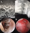
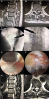
Background: Endoscopic spine surgery in the cervicothoracic spine is generating continued interest in a rapidly evolving field. The authors present 4 techniques for fully endoscopic cervical spine surgery: (1) posterior cervical unilateral laminectomy and bilateral decompression, (2) posterior cervical foraminotomy, (3) anterior cervical discectomy, and (4) anterior transcorporal discectomy. Two techniques for fully endoscopic thoracic spine surgery are also presented: (1) posterior thoracic unilateral laminectomy and bilateral decompression and (2) transforaminal thoracic endoscopic discectomy and foraminotomy.
Methods: We describe 6 different surgical approaches and review the relevant literature about each technique.
Results: The clinical application of endoscopic spine surgery techniques has evolved over the past 40 years. Recent data suggest comparable outcomes to other procedures and perhaps fewer complications and quicker recovery when these techniques are used in the cervical and thoracic spine. Significant variability exists in these approaches depending on the goal of canal decompression, root decompression, and the site of the pathology.
Conclusions: Each endoscopic approach in the cervicothoracic spine has its technical nuances, outcomes, advantages, and disadvantages, making fully endoscopic cervicothoracic spine surgery an exciting and growing field.
Keywords: TESSYS; cervical; endoscopic discectomy; radiculopathy; thoracic; transforaminal.
This manuscript is generously published free of charge by ISASS, the International Society for the Advancement of Spine Surgery. Copyright © 2021 ISASS. To see more or order reprints or permissions, see http://ijssurgery.com.
Conflict of interest statement
Declaration of Conflicting Interests: The authors report no conflicts of interest in this work.
Figures


Similar articles
-
Full endoscopic cervical spine surgery.J Spine Surg. 2020 Jun;6(2):383-390. doi: 10.21037/jss.2019.10.15. J Spine Surg. 2020. PMID: 32656375 Free PMC article.
-
Posterior endoscopic cervical foramiotomy and discectomy: clinical and radiological computer tomography evaluation on the bony effect of decompression with 2 years follow-up.Eur Spine J. 2021 Feb;30(2):534-546. doi: 10.1007/s00586-020-06637-8. Epub 2020 Oct 19. Eur Spine J. 2021. PMID: 33078265
-
Key Takeaways From the ISASS Webinar Series on Current and Emerging Techniques in Endoscopic Spine Surgery | Part 2: Polytomous Rasch Analysis of Learning Curve and Surgeon Endorsement of Biportal, Interlaminar, and Transforaminal Endoscopic Stenosis Decompression, Discectomy, and Laminectomy in Combination With Interspinous Process Spacers.Int J Spine Surg. 2024 Nov 20;18(S2):S23-S37. doi: 10.14444/8673. Int J Spine Surg. 2024. PMID: 39547677 Free PMC article.
-
Narrative Review of Uniportal Posterior Endoscopic Cervical Foraminotomy.World Neurosurg. 2024 Jan;181:148-153. doi: 10.1016/j.wneu.2023.10.021. Epub 2023 Oct 31. World Neurosurg. 2024. PMID: 37821026 Review.
-
Endoscopic Spine Surgery.J Korean Neurosurg Soc. 2017 Sep;60(5):485-497. doi: 10.3340/jkns.2017.0203.004. Epub 2017 Aug 30. J Korean Neurosurg Soc. 2017. PMID: 28881110 Free PMC article. Review.
Cited by
-
Transforaminal endoscopic thoracic discectomy: surgical technique.J Spine Surg. 2023 Jun 30;9(2):166-175. doi: 10.21037/jss-22-109. Epub 2023 Apr 13. J Spine Surg. 2023. PMID: 37435321 Free PMC article.
-
Full Endoscopic Surgery for Thoracic Pathology: Next Step after Mastering Lumbar and Cervical Endoscopic Spine Surgery?Biomed Res Int. 2022 May 16;2022:8345736. doi: 10.1155/2022/8345736. eCollection 2022. Biomed Res Int. 2022. PMID: 35615011 Free PMC article.
References
LinkOut - more resources
Full Text Sources
Miscellaneous
