

A case of retroperitoneal tumor displaying epithelial differentiation, prominent myxoid stroma and loss of INI1/SMARCB1
- PMID: 34974552
- PMCID: PMC8720391
- DOI: 10.32074/1591-951X-250
A case of retroperitoneal tumor displaying epithelial differentiation, prominent myxoid stroma and loss of INI1/SMARCB1
Abstract
The clinicopathological spectrum of INI1 deficient tumors is expanding. Epithelioid sarcoma (ES) is a rare sarcoma of uncertain differentiation, more often occurring in the extremities and uncommonly in the deep soft tissues. Histopathologically, it manifests in the form of classical, proximal, or hybrid types, the latter two characterized by rhabdoid cytomorphology. Immunohistochemically, ESs display loss of INI1/SMARCB1 and genetically associated with high percentage of SMARCB1 deletions.
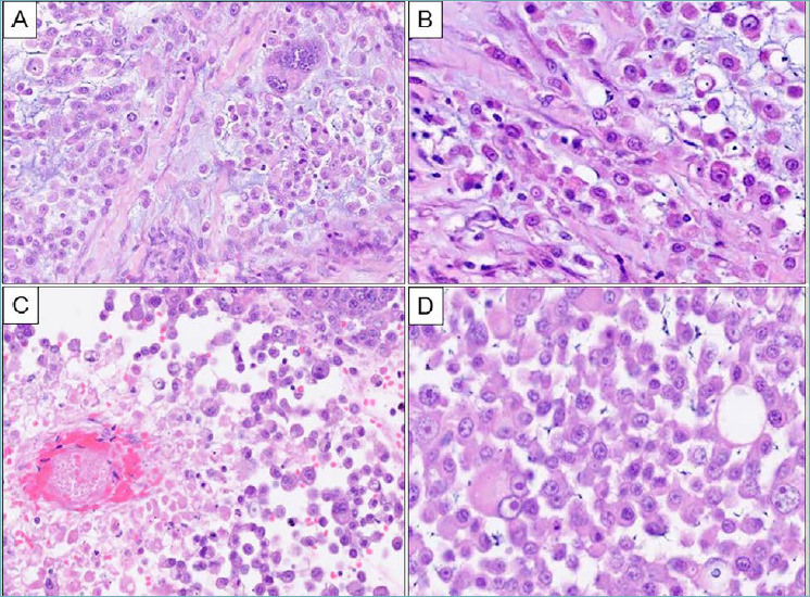
We report an extremely uncommon case of a retroperitoneal tumor in a 42-year-old male, who presented with abdominal discomfort. Radiologic imaging disclosed a 12 cm-sized retroperitoneal mass without involvement of any organ parenchyma. The patient underwent tumor excision with left-sided nephrectomy at another hospital. A review of the paraffin-embedded tissue sections revealed a multinodular tumor, composed of dyscohesive epithelioid tumor cells and focally arranged in cords, containing moderate to abundant, eosinophilic cytoplasm, vesicular nuclei, containing prominent nucleoli, including cells with rhabdoid cytomorphology, in a conspicuous myxoid stroma. A focal tumor area resembled proximal-type of ES. Immunohistochemically, tumor cells displayed positivity for pan cytokeratin (AE1/AE3), epithelial membrane antigen (EMA), vimentin and focally for CA125, while these were negative for CD34, S100 protein, CKIT, DOG1, and INI1/SMARCB1.
To the best of our knowledge, this constitutes the first case of a malignant tumor with epithelioid morphology, displaying myxoid matrix and loss of INI1/SMARCB1, resembling a myxoid variant of an epithelioid sarcoma and myoepithelioma-like tumor of the vulvar tumor, occurring in the retroperitoneum. A review of similar cases, differential diagnosis and treatment-associated implications are presented.
Keywords: INI1/SMARCB1; epithelioid sarcoma; myxoid tumors; rare tumor; retroperitoneal sarcoma.
Copyright © 2021 Società Italiana di Anatomia Patologica e Citopatologia Diagnostica, Divisione Italiana della International Academy of Pathology.
Conflict of interest statement
The Authors declare no conflict of interest.
Figures


References
-
- Hollmann TJ, Hornick JL. INI1-deficient tumors: diagnostic features and molecular genetics. Am J Surg Pathol 2011;35:e47-63. https://doi.org/10.1097/PAS.0b013e31822b325b 10.1097/PAS.0b013e31822b325b - DOI - PubMed
-
- Oda Y, Dal Cin P, Le Loarer F, Nielsen TO. Tumors of uncertain differentiation/Epithelioid sarcoma. In: World Health Organization (WHO) classification of tumours editorial board, eds. World Health Organization classification of tumours. 5th ed. Soft tissue and bone tumours. Lyon, France: IARC Press; 2020, pp. 294-296.
-
- Spillane AJ, Thomas JM, Fisher C. Epithelioid sarcoma: the clinicopathological complexities of this rare soft tissue sarcoma. Ann Surg Oncol 2000;7:218-225. https://doi.org/10.1007/BF02523657 10.1007/BF02523657 - DOI - PubMed
-
- Fisher C. Epithelioid sarcoma of Enzinger. Adv Anat Pathol 2006;13:114-121. https://doi.org/10.1097/00125480-200605000-00002 10.1097/00125480-200605000-00002 - DOI - PubMed
-
- Rekhi B, Gorad BD, Chinoy RF. Clinicopathological features with outcomes of a series of conventional and proximal-type epithelioid sarcomas, diagnosed over a period of 10 years at a tertiary cancer hospital in India. Virchows Arch 2008;453:141-153. https://doi.org/10.1007/s00428-008-0639-0 10.1007/s00428-008-0639-0 - DOI - PubMed
Publication types
MeSH terms
Substances
LinkOut - more resources
Full Text Sources
Medical
Research Materials
Miscellaneous
