. 2022 Jan 1;18(1):112-123.
doi: 10.7150/ijbs.63388. eCollection 2022.

Rab31 promotes the invasion and metastasis of cervical cancer cells by inhibiting MAPK6 degradation

Affiliations

Rab31 promotes the invasion and metastasis of cervical cancer cells by inhibiting MAPK6 degradation

Yujie Huang et al. Int J Biol Sci. .

Abstract

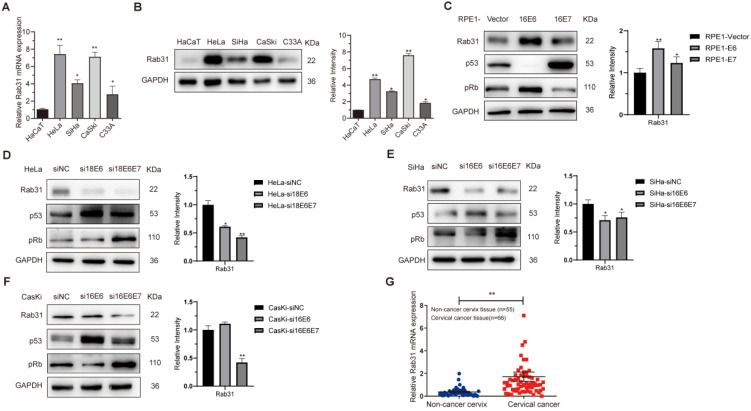
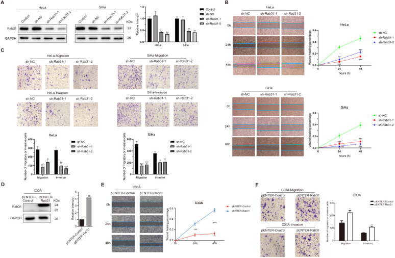
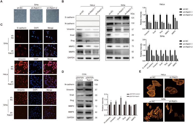
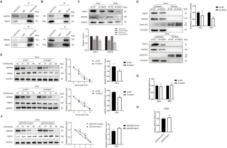
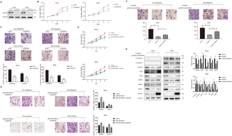
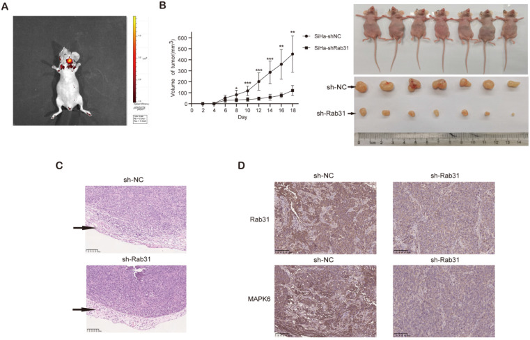
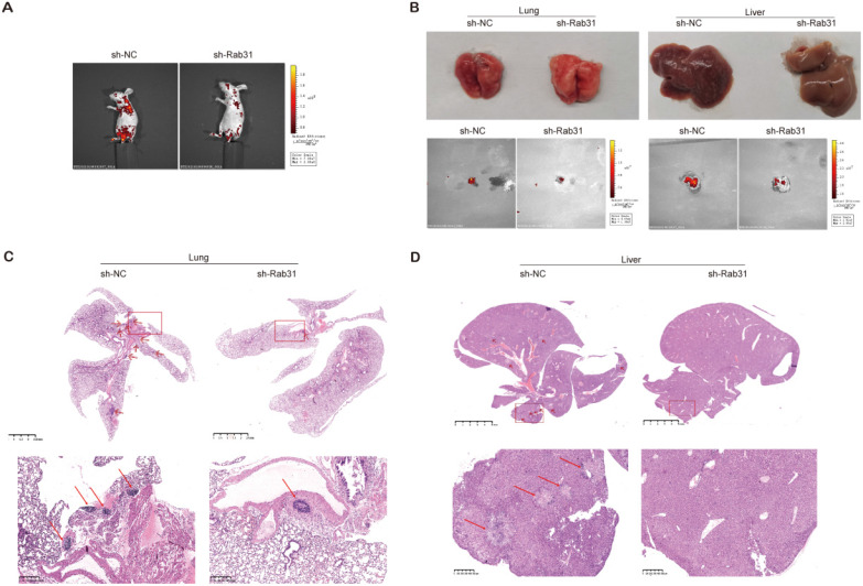
Persistent infection with high-risk human papillomavirus (HPV) is the main risk factor for cervical cancer. Our mass spectrometry data showed that the Ras-associated binding protein Rab31 was upregulated by HPV; however, little is known regarding the role of Rab31 in the metastasis of cervical cancer cells. In this study, we showed that Rab31 was highly expressed in cervical cancer tissues and cells, and both HPV E6 and E7 promoted the expression of Rab31. Rab31 knockdown inhibited while Rab31 overexpression promoted the migration and invasion capabilities of cervical cancer cells. Additionally, Rab31 knockdown inhibited the epithelial-mesenchymal transition (EMT) and cytoskeletal rearrangement in cervical cancer cells. Furthermore, Rab31 interacted with mitogen-activated protein kinase 6 (MAPK6), and Rab31 knockdown inhibited the expression of MAPK6, which was mainly localized in the cytoplasm. More importantly, Rab31 knockdown promoted and Rab31 overexpression inhibited MAPK6 degradation. Accordingly, MAPK6 overexpression restored the decreased migration potential caused by Rab31 knockdown. Finally, a xenograft mouse model showed that Rab31 knockdown in cervical cancer cells led to reduced tumor growth and impaired lung and liver metastasis in vivo. In conclusion, Rab31 plays a crucial role in cervical cancer metastasis by inhibiting MAPK6 degradation. Thus, Rab31 may serve as a novel therapeutic target to manage cervical cancer.

Keywords: Cervical cancer; HPV; MAPK6; Metastasis; Rab31.


Conflict of interest statement

Competing Interests: The authors have declared that no competing interest exists.

Figures

Figure 1
Figure 1
Rab31 is highly expressed in cervical cancer cells and tissues. (A) qRT-PCR was used to detect the mRNA level of Rab31 in different cells, and the mRNA level of Rab31 was higher in cervical cancer cells than in non-cancer HaCaT cells. (B) Rab31 protein level was detected by Western blotting, and Rab31 was highly expressed in cervical cancer cells. (C) Western blotting of Rab31, p53 and pRb in HPV16 E6- and E7-expressing RPE1 cells was shown. (D-F) Detection of Rab31, p53 and pRb protein levels in HeLa, SiHa and CaSki cells after transfection with siRNA targeting HPV18 E6, 18 E6E7 or 16 E6, 16 E6E7. (G) Detection of Rab31 mRNA level in 55 non-cancer cervical tissues and 66 cervical cancer tissue specimens using qRT-PCR, and Rab31 was highly expressed in cervical cancer tissues. *P <0.05, **P <0.01.
Figure 2
Figure 2
Rab31 promotes the migration and invasion of cervical cancer cells. (A) Two shRNA lentiviruses targeting Rab31 were used to infect HeLa and SiHa cervical cancer cells, and Western blotting showed that Rab31 was efficiently knocked down. (B) A wound-healing assay was used to detect the capability of cancer cells to migrate at 0 h, 24 h and 48 h. The statistical graph on the right is a quantitative analysis of the wound-healing measured at different time points. (C) The effect of Rab31 knockdown on cell migration and invasion was analyzed by Transwell assay. The pictures below showed the statistical results. (D) The Rab31 expression plasmid was transfected into C33A cells, and Rab31 expression was detected by Western blotting. (E) A wound-healing assay was performed and detected at 0 h, 24 h and 48 h. (F) The effect of Rab31 overexpression on cell migration and invasion was explored using Transwell assay. *P <0.05, **P <0.01, ***P <0.001.
Figure 3
Figure 3
Rab31 promotes the migration and invasion via EMT and cytoskeletal rearrangement. (A) The effect of Rab31 knockdown on the morphology of SiHa cells was shown. (B) Western blotting of the protein levels of epithelial marker E-cadherin, mesenchymal markers (N-cadherin, Vimentin), EMT transcription factors Snail and Slug as well as MMP2, MMP9 in Rab31-knockdown HeLa and SiHa cells were shown. (C) Cellular immunofluorescence assays were performed to detect the expression of E-cadherin and Vimentin in HeLa and SiHa cells. (D) Expressions of EMT-related proteins in Rab31-overexpressing C33A cells were measured. (E) The filamentous actin backbone of HeLa and SiHa cells was fluorescently stained with phalloidin, and the cytoskeletal structure was observed under a fluorescence microscope. The arrow in the picture above indicates F-actin and the arrow in the picture below indicates the pseudopod. *P <0.05, **P <0.01, ***P <0.001.
Figure 4
Figure 4
Rab31 inhibits MAPK6 degradation. (A) The interaction of endogenous Rab31 and MAPK6 in SiHa cells was detected using the Co-IP assay. (B) Transfection of plasmids expressing Rab31 (Flag tag) and MAPK6 (HA tag) into SiHa cells and the interaction between exogenous Rab31 and MAPK6 was detected. (C) Detection of MAPK6 protein expression after Rab31 knockdown. (D) Western blotting of MAPK6 cellular localization following Rab31 knockdown was shown. (E) Cells with Rab31 knockdown were incubated with 5 μg/mL of CHX and harvested at the indicated times. MAPK6 stability was monitored by the line chart, and the half-life of MAPK6 was evaluated on the right. (F) Cells with Rab31 overexpression were incubated with 5 μg/mL of CHX and harvested at the indicated times. MAPK6 stability and the half-life of MAPK6 were shown. (G-H) The mRNA level of MAPK6 was detected following Rab31 knockdown or Rab31 overexpression using qRT-PCR. *P <0.05, **P <0.01, ***P <0.001.
Figure 5
Figure 5
Rab31 promotes the invasion of cervical cancer cells partly via MAPK6. (A) Detection of MAPK6 protein in HeLa and SiHa cells after MAPK6 knockdown. (B) MAPK6 was knocked down in HeLa and SiHa cells, and CCK-8 assay was performed. (C) The migration and invasion of HeLa and SiHa cells were tested by Transwell assay following MAPK6 knockdown, and the pictures below show the statistical results. (D) The viability of Rab31-overexpressing cells following MAPK6 knockdown was analyzed by the CCK-8 assay. The significant difference of viability between Rab31-overexpressing cells with MAPK6 knockdown and Rab31-overexpressing cells was marked with #. (E) The migration and invasion of Rab31-overexpressing cells following MAPK6 knockdown were tested by Transwell assay. The pictures on the right show the statistical results. (F) The effect of MAPK6 overexpression on the metastatic capability of Rab31-knockdown HeLa and SiHa cells was detected using Transwell assay and the pictures below show the statistical results. (G) EMT-related proteins in Rab31-knockdown cells with MAPK6 overexpression were detected by Western blotting. *P <0.05, **P <0.01, ***P <0.001, #P <0.05, ##P <0.01, ###P <0.001.
Figure 6
Figure 6
Rab31 knockdown inhibits tumor growth in nude mice. (A) In vivo fluorescence imaging of nude mice showed the fluorescence intensity of the tumors in the left and right flanks. (B) Tumor growth was monitored, and the volume was measured every 2 days with calipers. The growth curve of tumors was shown (left). The nude mice and tumors were photographed after the nude mice were euthanized (right). (C) H&E staining of transplanted tumor was used to detect the integrity of the tumor envelope. The arrows indicate the tumor envelope. (D) Immunohistochemical staining of Rab31 and MAPK6 in transplanted tumors was shown. *P<0.05, **P <0.01, ***P<0.001.
Figure 7
Figure 7
Rab31 knockdown inhibits the metastasis of cervical cancer cells in vivo. (A) In vivo fluorescence imaging of nude mice showed that fluorescence was mainly located in the chest and abdomen. (B) Visible lung and liver metastatic nodules and fluorescence imaging of the lung and liver were shown. (C-D) H&E staining of paraffin-embedded sections of lung and liver metastatic nodules was shown. The pictures below are enlargements of the rectangle in the pictures above. The arrows indicate the metastatic nodules.

References

    1. Burd EM. Human papillomavirus and cervical cancer. CLIN MICROBIOL REV. 2003;16:1–17. - PMC - PubMed
    1. Munoz N, Bosch FX, de Sanjose S, Herrero R, Castellsague X, Shah KV. et al. Epidemiologic classification of human papillomavirus types associated with cervical cancer. N Engl J Med. 2003;348:518–27. - PubMed
    1. Moody CA, Laimins LA. Human papillomavirus oncoproteins: pathways to transformation. NAT REV CANCER. 2010;10:550–60. - PubMed
    1. Dyson N, Howley PM, Munger K, Harlow E. The human papilloma virus-16 E7 oncoprotein is able to bind to the retinoblastoma gene product. SCIENCE. 1989;243:934–7. - PubMed
    1. Chellappan S, Kraus VB, Kroger B, Munger K, Howley PM, Phelps WC. et al. Adenovirus E1A, simian virus 40 tumor antigen, and human papillomavirus E7 protein share the capacity to disrupt the interaction between transcription factor E2F and the retinoblastoma gene product. Proc Natl Acad Sci U S A. 1992;89:4549–53. - PMC - PubMed

Publication types

Substances