. 2021 Dec 16:15:782786.
doi: 10.3389/fnins.2021.782786. eCollection 2021.

Self-Administration of Right Vagus Nerve Stimulation Activates Midbrain Dopaminergic Nuclei

Affiliations

Self-Administration of Right Vagus Nerve Stimulation Activates Midbrain Dopaminergic Nuclei

Jackson Brougher et al. Front Neurosci. .

Abstract

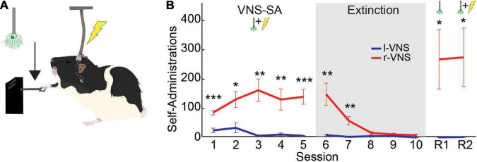
Background: Left cervical vagus nerve stimulation (l-VNS) is an FDA-approved treatment for neurological disorders including epilepsy, major depressive disorder, and stroke, and l-VNS is increasingly under investigation for a range of other neurological indications. Traditional l-VNS is thought to induce therapeutic neuroplasticity in part through the coordinated activation of multiple broadly projecting neuromodulatory systems in the brain. Recently, it has been reported that striking lateralization exists in the anatomical and functional connectivity between the vagus nerves and the dopaminergic midbrain. These emerging findings suggest that VNS-driven activation of this important plasticity-promoting neuromodulatory system may be preferentially driven by targeting the right, rather than the left, cervical nerve. Objective: To compare the effects of right cervical VNS (r-VNS) vs. traditional l-VNS on self-administration behavior and midbrain dopaminergic activation in rats. Methods: Rats were implanted with a stimulating cuff electrode targeting either the right or left cervical vagus nerve. After surgical recovery, rats underwent a VNS self-administration assay in which lever pressing was paired with r-VNS or l-VNS delivery. Self-administration was followed by extinction, cue-only reinstatement, and stimulation reinstatement sessions. Rats were sacrificed 90 min after completion of behavioral training, and brains were removed for immunohistochemical analysis of c-Fos expression in the dopaminergic ventral tegmental area (VTA) and substantia nigra pars compacta (SNc), as well as in the noradrenergic locus coeruleus (LC). Results: Rats in the r-VNS cohort performed significantly more lever presses throughout self-administration and reinstatement sessions than did rats in the l-VNS cohort. Moreover, this appetitive behavioral responding was associated with significantly greater c-Fos expression among neuronal populations within the VTA, SNc, and LC. Differential c-Fos expression following r-VNS vs. l-VNS was particularly prominent within dopaminergic midbrain neurons. Conclusion: Our results support the existence of strong lateralization within vagal-mesencephalic signaling pathways, and suggest that VNS targeted to the right, rather than left, cervical nerve preferentially activates the midbrain dopaminergic system. These findings raise the possibility that r-VNS could provide a promising strategy for enhancing dopamine-dependent neuroplasticity, opening broad avenues for future research into the efficacy and safety of r-VNS in the treatment of neurological disease.

Keywords: VNS (vagus nerve stimulation); c-fos; dopamine; lateralization; neural stimulation; self-administration; substantia nigra; ventral tegmental area.


Conflict of interest statement

The authors declare that the research was conducted in the absence of any commercial or financial relationships that could be construed as a potential conflict of interest.

Figures

FIGURE 1
FIGURE 1
Rats self-administer r-VNS but not l-VNS. (A) During VNS-SA sessions, r-VNS or l-VNS was delivered along with a visual cue (488 nm LED) immediately upon detection of a lever deflection. (B) Lever-press performance of r-VNS (red; n = 7) and l-VNS (blue; n = 7) cohorts throughout the self-administration assay. Training stages included VNS self-administration (VNS-SA), Extinction, cue-only reinstatement (R1), and yoked-stimulation reinstatement (R2) sessions. Student’s t-tests were used to test for between-group differences in behavioral performance within each training session and corrected for multiple comparisons using false discovery rate. For sessions in which FDR-corrected comparisons indicated the presence of a statistically significant between-group difference, uncorrected p-values are denoted: *p < 0.05, **p < 0.01, ***p < 0.001.
FIGURE 2
FIGURE 2
Compared to l-VNS, r-VNS self-administration increases c-Fos expression within catecholaminergic nuclei. (A,C,E) Representative 10x images and ROI boundaries used to quantify c-Fos expression within the VTA (A), SNc (C), and LC (E) following either l-VNS (left) or r-VNS (right). Sections were stained for tyrosine hydroxylase (red) to label catecholaminergic neurons, c-Fos (green) as a marker of neuronal activation, and the nuclear marker DAPI (omitted for clarity). (B,D,F) Mean gray value (MGV) of c-Fos fluorescence was significantly greater following r-VNS self-administration (red) than in l-VNS treated rats (blue) within both left (L) and right (R) brain hemispheres of the VTA (B), SNc (D), and LC (F). **p < 0.01; ****p < 0.0001; 2-way ANOVA between-group comparisons. Within each treatment group, no significant difference in c-Fos expression was observed between left and right brain hemispheres; full statistical results are presented in Tables 2, 3.
FIGURE 3
FIGURE 3
Compared to l-VNS, r-VNS self-administration significantly increases c-Fos expression in both TH+ and TH− cells within catecholaminergic nuclei. (A,C,E) Representative 20x images and ROI boundaries used to quantify single-cell c-Fos expression within the VTA (A), SNc (C), and LC (E) following either l-VNS (left) or r-VNS (right). Sections were co-stained for tyrosine hydroxylase (red), c-Fos (green), and DAPI (blue). Arrow heads in enlarged insets show example cells classified as exclusively DAPI+ (cyan arrows); DAPI+, c-Fos+, and TH− (green arrows); DAPI+, c- Fos-, and TH+ (magenta arrows); or DAPI+, c-Fos+, and TH+ (white arrows). (B,D,F) In both VTA (B) and SNc (D), the percentage of TH+ neurons did not differ between r-VNS and l-VNS treatment groups (top). However, the percentage of c-Fos+ cells (bottom) was significantly greater in the r-VNS group, in both TH+ and TH− cell populations. (F) In the LC, r-VNS self-administration resulted in a higher percentage of TH+ cells than l-VNS (top), but the percentage of TH+ and TH− cells that were found to be c-Fos+ did not differ between groups (bottom). In (B,D,F), Student’s t-tests were used to test for between-group differences in TH+ population size, as well as in c-Fos expression within TH+ and TH− populations; multiple comparisons were corrected using false discovery rate. For FDR-corrected comparisons in which statistically significant differences were observed, uncorrected p-values are indicated: *p < 0.05, **p < 0.01, ***p < 0.001.

References

    1. Anand I. S., Konstam M. A., Klein H. U., Mann D. L., Ardell J. L., Gregory D. D., et al. (2020). Comparison of symptomatic and functional responses to vagus nerve stimulation in ANTHEM-HF. INOVATE-HF, and NECTAR-HF. ESC Heart Fail. 7 75–83. 10.1002/ehf2.12592 - DOI - PMC - PubMed
    1. Ardell J. L., Randall W. C. (1986). Selective vagal innervation of sinoatrial and atrioventricular nodes in canine heart. Am. J. Physiol. 251(4 Pt 2) H764–H773. 10.1152/ajpheart.1986.251.4.h764 - DOI - PubMed
    1. Bai L., Mesgarzadeh S., Ramesh K. S., Huey E. L., Liu Y., Gray L. A., et al. (2019). Genetic identification of vagal sensory neurons that control feeding. Cell 179 1129.e23–1143.e23. 10.1016/j.cell.2019.10.031 - DOI - PMC - PubMed
    1. Baik J. H. (2013). Dopamine signaling in reward-related behaviors. Front. Neural Circuits 7:152. 10.3389/fncir.2013.00152 - DOI - PMC - PubMed
    1. Berthoud H. R. (2008). Vagal and hormonal gut-brain communication: from satiation to satisfaction. Neurogastroenterol. Motil. 20 64–72. 10.1111/j.1365-2982.2008.01104.x - DOI - PMC - PubMed

LinkOut - more resources