. 2021 Dec 16:12:774316.
doi: 10.3389/fphar.2021.774316. eCollection 2021.

The Preventive Effect of Decorin on Epidural Fibrosis and Epidural Adhesions After Laminectomy

Affiliations

The Preventive Effect of Decorin on Epidural Fibrosis and Epidural Adhesions After Laminectomy

Qing Ding et al. Front Pharmacol. .

Abstract

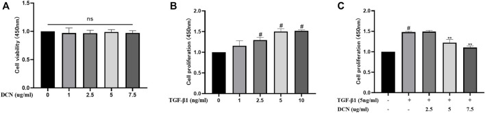
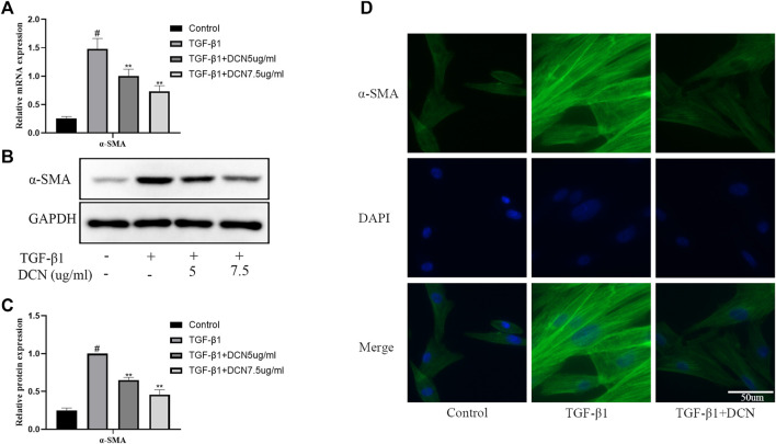
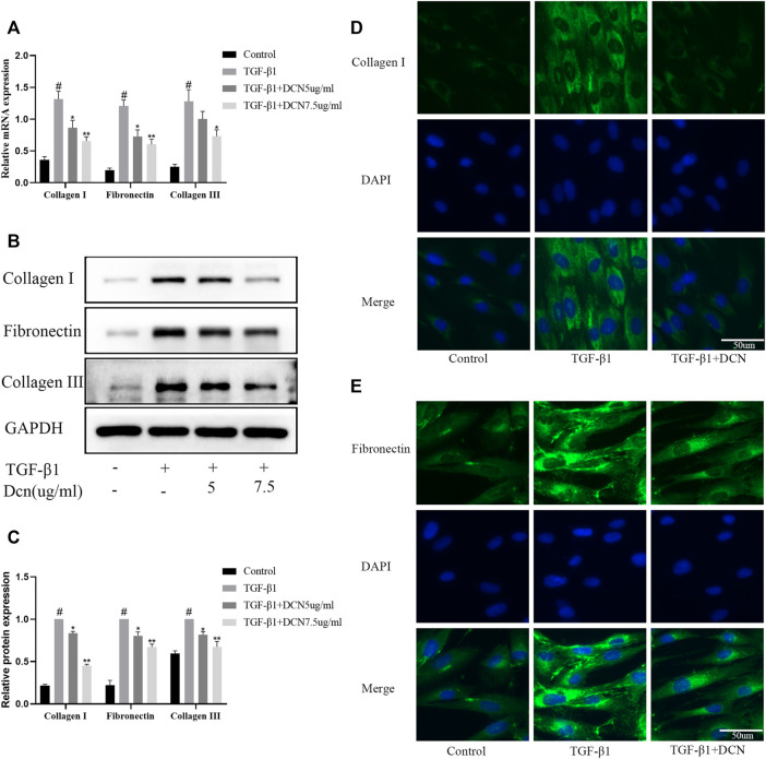
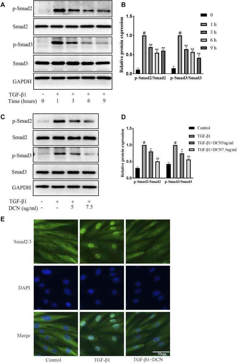
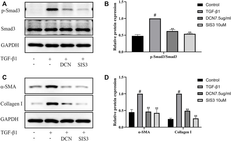
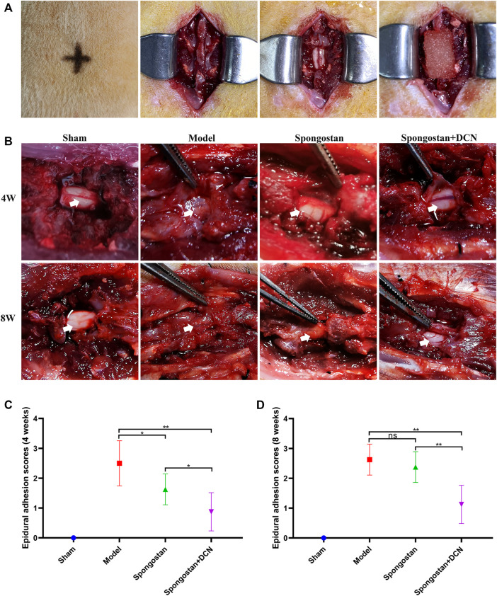
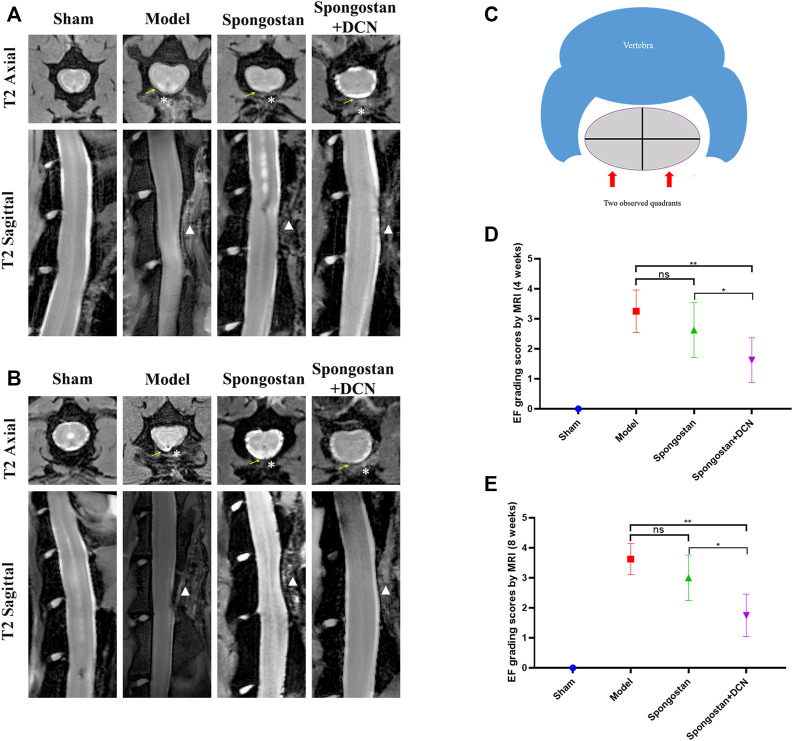
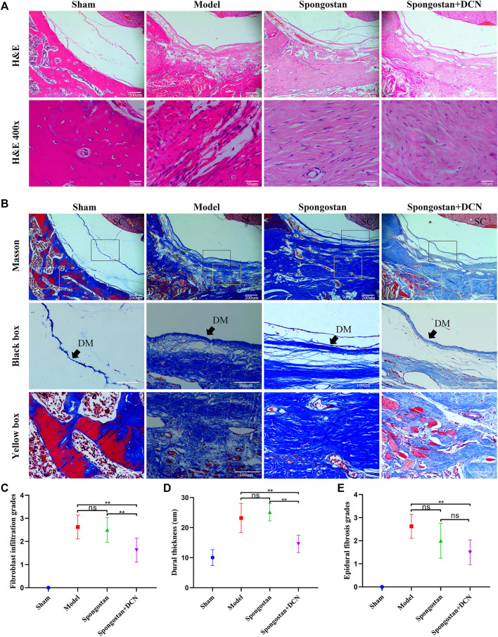
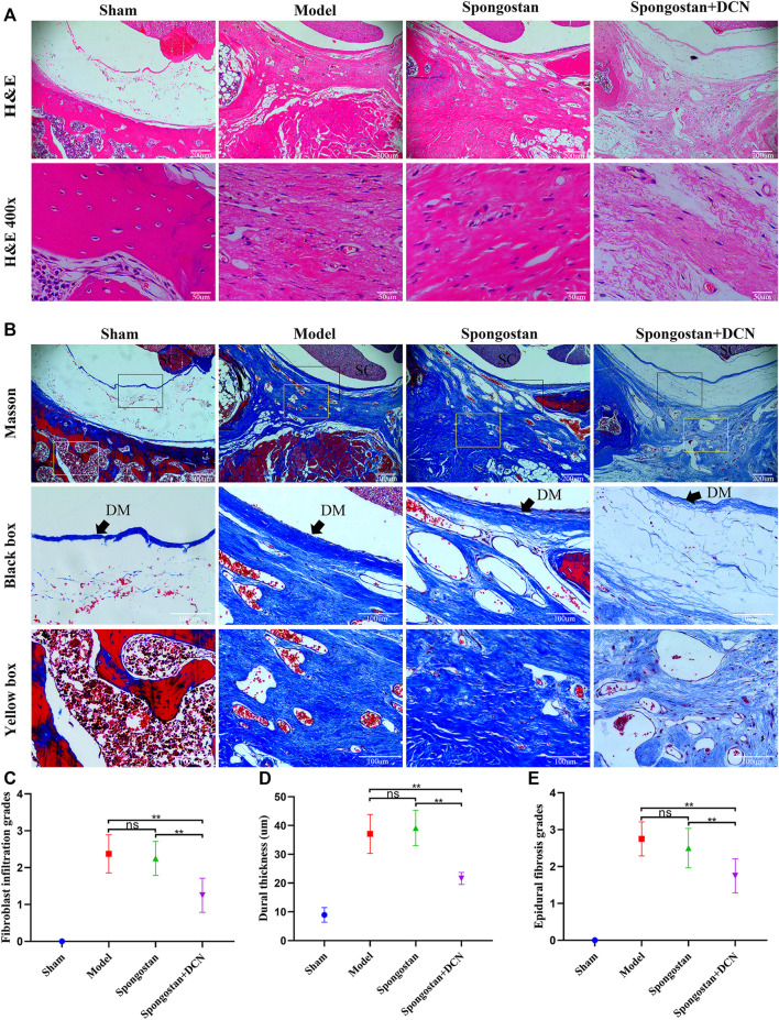
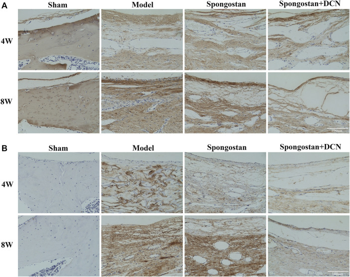
Laminectomy is commonly performed to treat degenerative spinal diseases by reducing compression on the spinal cord and nerve roots. The postoperative epidural fibrosis and epidural adhesions may result in failed back surgery syndrome, which is characterized by the symptoms of lower back pain or leg pain. There is currently no satisfactory treatment for this complication. The pathological processes of epidural fibrosis and epidural adhesions are relevant to the proliferation of fibroblasts, transdifferentiation of fibroblasts into myofibroblasts, and the excessive deposition of extracellular matrix (ECM) protein. According to reports, transforming growth factor-β1 (TGF-β1) played a vital role in the development of fibrosis by promoting aforementioned processes. Decorin, an endogenous proteoglycan and natural inhibitor of TGF-β1, has exhibited prominent anti-fibrosis activity in various scar formation and fibrosis models of many organs. However, the preventive effect of decorin on epidural fibrosis and epidural adhesions requires further investigation. Here, we investigated the therapeutic effects and potential mechanisms of decorin on epidural fibrosis and epidural adhesions. Our results indicated that decorin could significantly suppress the TGF-β1-induced proliferation, transdifferentiation, and extracellular matrix production in primary fibroblasts. Furthermore, Smad2/3 signaling pathway had been demonstrated to be involved in the preventive effect of decorin. Moreover, administration of decorin in vivo could notably inhibit epidural fibrosis and epidural adhesions after laminectomy. To date, there is no approved therapy to target TGF-β1 for the treatment of epidural fibrosis and epidural adhesions after laminectomy. Our research proved the anti-fibrosis effect of decorin, which may provide an effective and promising treatment for epidural fibrosis and epidural adhesions.

Keywords: Smad2/3; decorin; epidural adhesion; epidural fibrosis; fibroblast; laminectomy.


Conflict of interest statement

The authors declare that the research was conducted in the absence of any commercial or financial relationships that could be construed as a potential conflict of interest.

Figures

FIGURE 1
FIGURE 1
Effects of decorin (DCN) on cell viability and TGF-β1-induced proliferation are evaluated using a CCK-8 kit. The absorbance of the cells at 450 nm from each group is detected and recorded. (A) Fibroblasts were exposed to DCN (1, 2.5, 5, and 7.5 µg/ml) or (B) TGF-β1 (1, 2.5, 5, and 10 ng/ml) for 48 h. (C) Fibroblasts were treated with TGF-β1 (5 ng/ml) with or without DCN (2.5, 5, and 7.5 µg/ml) for 48 h. ns indicates no significance. # p < 0.05 vs control group; **p < 0.01 vs TGF-β1 group.
FIGURE 2
FIGURE 2
Decorin (DCN) suppresses TGF-β1-induced transdifferentiation of fibroblasts. Fibroblasts were exposed to TGF-β1 (5 ng/ml) with or without DCN (5 and 7.5 µg/ml) for 48 h. (A) Gene expression of α-SMA was detected by RT-qPCR. (B) Western blotting and (C) quantitative analysis of α-SMA in each group. (D) The α-SMA was observed by immunofluorescence after cells were treated with TGF-β1 (5 ng/ml) with or without DCN (7.5 µg/ml). # p < 0.05 vs control group; **p < 0.01 vs TGF-β1 group.
FIGURE 3
FIGURE 3
Decorin (DCN) inhibits TGF-β1-induced extracellular matrix synthesis. Fibroblasts were exposed to TGF-β1 (5 ng/ml) with or without DCN (5 and 7.5 µg/ml) for 48 h. (A) Gene expression of fibronectin, collagen I, and collagen III was detected by RT-qPCR. (B) Western blotting and (C) quantitative analysis of fibronectin, collagen I, and collagen III in each group. (D) Collagen I and (E) fibronectin were observed by immunofluorescence after cells were treated with TGF-β1 (5 ng/ml) with or without DCN (7.5 µg/ml). # p < 0.05 vs control group; *p < 0.05 vs TGF-β1 group; **p < 0.01 vs TGF-β1 group.
FIGURE 4
FIGURE 4
Decorin (DCN) inhibits TGF-β1-induced Smad2/3 signaling pathway activation. (A) Western blotting and (B) quantification analysis of Smad2, p-Smad2, Smad3, and p-Smad3 from TGF-β1-treated fibroblasts at different time points (0, 1, 3, 6, and 9 h). (C) Western blotting and (D) quantification analysis of Smad2, p-Smad2, Smad3, and p-Smad3 from fibroblasts after treated with DCN (5 and 7.5 µg/ml) for 2 h, followed by incubation with TGF-β1 (5 ng/ml) for 1 h. (E) Nuclear translocation of Smad2/3 was detected by immunofluorescence. # p < 0.05 vs control group; *p < 0.05 vs TGF-β1 group; **p < 0.01 vs TGF-β1 group.
FIGURE 5
FIGURE 5
TGF-β1-induced Smad2/3 signaling pathway activation, fibroblast transdifferentiation, and ECM synthesis are inhibited by SIS3 (a novel specific inhibitor of Smad3). (A) Western blotting and (B) quantification analysis of Smad3 and p-Smad3 from fibroblasts after treated with DCN (7.5 µg/ml) or SIS3 (10 µM) for 2 h, followed by incubation with TGF-β1 (5 ng/ml) for 1 h. (C) Western blotting and (D) quantification analysis of α-SMA and collagen I from fibroblasts after treated with TGF-β1 (5 ng/ml) in the presence of DCN (7.5 µg/ml) or SIS3 (10 µM) for 48 h. # p < 0.05 vs control group; *p < 0.05 vs TGF-β1 group; **p < 0.01 vs TGF-β1 group.
FIGURE 6
FIGURE 6
Establishment of a rat laminectomy model and gross observation of the epidural adhesions. (A) The process of building the model: surface location of L1, exposing the lamina, excision of lamina, and a spongostan impregnated with saline or decorin (DCN) solution was retained on the dura mater. (B) Gross observation of laminectomy sites after 4 and 8 weeks of treatment. White arrows indicate the dura mater. (C) Four weeks and (D) 8 weeks of epidural adhesion score results based on Rydell standard. ns indicates no significance. *p < 0.05; **p < 0.01.
FIGURE 7
FIGURE 7
Results of MRI evaluation. The axial and sagittal T2-weighted MRIs of laminectomy sites at (A) 4 weeks and (B) 8 weeks. Yellow arrows indicate the dura mater. * indicates the epidural dense scar tissues. White triangles in sagittal images indicate lamina defect site. (C) Schematic diagram of the axial image used for epidural fibrosis (EF) scoring. (D) Four weeks and (E) 8 weeks EF score results based on MRIs. ns indicates no significance. *p < 0.05; **p < 0.01.
FIGURE 8
FIGURE 8
Representative H&E staining and Masson staining from each group at 4 weeks postoperatively. (A) H&E staining of spinal column in laminectomy sites. The high-resolution images (400×) in the bottom panel show fibroblast infiltration. (B) Masson-stained sections of laminectomy sites. DM, dura mater; SC, spinal cord. (C) Quantified fibroblast infiltration grades at high resolution (400×). (D) Comparison of dural thickness among groups. (E) Quantitative analysis of epidural fibrosis grades according to Masson-stained sections. *p < 0.05; **p < 0.01.
FIGURE 9
FIGURE 9
Representative H&E staining and Masson staining from each group at 8 weeks postoperatively. (A) H&E staining of spinal column in laminectomy sites. The high-resolution images (400×) in the bottom panel show fibroblast infiltration. (B) Masson-stained sections of laminectomy sites. DM, dura mater; SC, spinal cord. (C) Quantified fibroblast infiltration grades at high resolution (400×). (D) Comparison of dural thickness among groups. (E) Quantitative analysis of epidural fibrosis grades according to Masson-stained sections. *p < 0.05; **p < 0.01.
FIGURE 10
FIGURE 10
Decorin (DCN) inhibits the synthesis of collagen I and fibronectin in vivo. Immunohistochemical staining of collagen I (A) and fibronectin (B) from each group at 4 and 8 weeks, respectively.

References

    1. Ahmed Z., Bansal D., Tizzard K., Surey S., Esmaeili M., Gonzalez A. M., et al. (2014). Decorin Blocks Scarring and Cystic Cavitation in Acute and Induces Scar Dissolution in Chronic Spinal Cord Wounds. Neurobiol. Dis. 64, 163–176. 10.1016/j.nbd.2013.12.008 - DOI - PubMed
    1. Alan C., Kocoglu H., Altintas R., Alici B., Resit Ersay A. (2011). Protective Effect of Decorin on Acute Ischaemia-Reperfusion Injury in the Rat Kidney. Arch. Med. Sci. 7 (2), 211–216. 10.5114/aoms.2011.22069 - DOI - PMC - PubMed
    1. Border W. A., Noble N. A., Yamamoto T., Harper J. R., Yamaguchi Yu., Pierschbacher M. D., et al. (1992). Natural Inhibitor of Transforming Growth Factor-Beta Protects against Scarring in Experimental Kidney Disease. Nature 360 (6402), 361–364. 10.1038/360361a0 - DOI - PubMed
    1. Braun K. M., Diamond M. P. (2014). The Biology of Adhesion Formation in the Peritoneal Cavity. Semin. Pediatr. Surg. 23 (6), 336–343. 10.1053/j.sempedsurg.2014.06.004 - DOI - PubMed
    1. Chan C. W., Peng P. (2011). Failed Back Surgery Syndrome. Pain Med. 12 (4), 577–606. 10.1111/j.1526-4637.2011.01089.x - DOI - PubMed

LinkOut - more resources