Case Reports
. 2021 Dec 15;17(3):514-520.
doi: 10.1016/j.radcr.2021.11.033. eCollection 2022 Mar.

Trans-scaphoid lunate dislocation: A case series

Affiliations
Case Reports

Trans-scaphoid lunate dislocation: A case series

Albert Gjeluci et al. Radiol Case Rep. .

Erratum in

Abstract

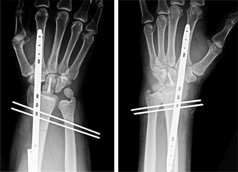
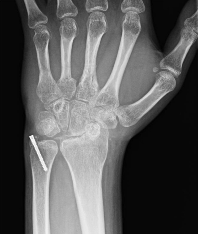
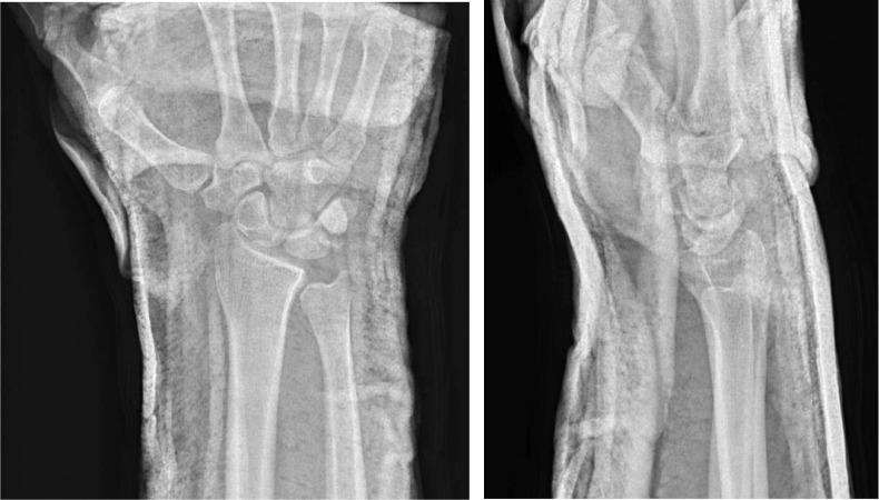
Trans-scaphoid lunate dislocation with volar displacement into the wrist/distal forearm is a devastating injury that most commonly occurs under situations of forceful impact to an extended wrist. Due to ligamentous disruption as well as fragile blood supply, these Mayfield type 4 injuries are associated with significant morbidity and long-term sequelae. Current treatment approaches to lunate dislocations depend on the severity and chronicity of the injury in addition to patient factors, with operative management potentially including ORIF or proximal row carpectomy. We report 5 cases of this rare injury pattern in 4 different patients.

Keywords: AVN, avascular necrosis; Avascular necrosis; Carpal instability; DRUJ, distal radioulnar joint; Lunate dislocation; Mayfield classification; ORIF, open reduction internal fixation; Perilunate dislocation; Perilunate instability; TFCC, triangular fibrocartilage complex.


Figures

Fig 1 –
Fig 1a
Patient 1: Initial radiographs of the right wrist showing a trans-scaphoid lunate dislocation. There is a fracture of the scaphoid waist with the proximal pole displaced into the volar wrist. There is severe dislocation of the lunate into the distal volar forearm.
Fig 1 –
Fig 1b
Patient 1: Postoperative radiograph of the right wrist showing changes of proximal row carpectomy.
Fig 2 –
Fig 2a
Patient 2: Initial radiographs of the right wrist demonstrate trans-capitate trans-scaphoid lunate dislocation as well as Galeazzi type fracture-dislocation including positive ulnar variance.
Fig 2 –
Fig 2b
Patient 2: Initial radiographs of the left wrist demonstrate trans-ulnar styloid trans-capitate trans-scaphoid lunate dislocation. Overlying splint material is present.
Fig 2 –
Fig 2c
Patient 2: Postoperative radiographs of the bilateral wrists demonstrate proximal row carpectomy, fixation of the capitate with a headless screw, radiocarpal fixation with a long dorsal plate that spans the distal radius and index metacarpal, and percutaneous pinning of the DRUJ. Postoperative findings are similar bilaterally with the exception of partially imaged plate and screw fixation of the radial diaphysis on the right and a nondisplaced ulnar styloid fracture on the left.
Fig 3 –
Fig 3a
Patient 3: Initial radiographs of the left wrist demonstrating a trans-ulnar styloid trans-scaphoid lunate dislocation. A nondisplaced triquetral fracture was noted on subsequent CT. There are also fractures of the third and fourth metacarpal bases.
Fig 3 –
Fig 3b
Patient 3: Postoperative radiographs of the left wrist demonstrating pinning across the carpal bones and the third and fourth metacarpal fractures, and screw fixation of the ulnar styloid fracture with improved alignment. Overlying splint material is present.
Fig 3 –
Fig 3c
Patient 3: Subsequent radiographs obtained 7 months after the radiographs in Figure 3b show interval removal of hardware, with a headless screw remaining across the ulnar styloid fracture. The proximal pole of the scaphoid and the lunate are no longer visible, compatible with bony resorption/collapse; these bones were not surgically removed. The capitate now articulates with the distal radius with associated degenerative changes. The third and fourth metacarpal base fractures demonstrate interval healing.
Fig 4 –
Fig 4a
Patient 4: Initial radiographs of the right wrist reveal a trans-radial styloid trans-scaphoid lunate dislocation. Irregular fracture fragments at the ulnar volar aspect of the wrist may be from the scaphoid or pisiform. The radial styloid fracture is nondisplaced. There is negative ulnar variance.
Fig 4 –
Fig 4b
Patient 4: Subsequent radiographs of the right wrist following reduction and splinting demonstrating improved alignment.

References

    1. Mayfield JK, Johnson RP, Kilcoyne RK. Carpal dislocations: pathomechanics and progressive perilunar instability. J Hand Surg Am. 1980;5(3):226–241. doi: 10.1016/s0363-5023(80)80007-4. - DOI - PubMed
    1. Arango D, Tiedeken NC, Ayzenberg M, Raphael J. Open perilunate injury with lunate revascularization after complete ligamentous avulsion. J Surg Case Rep. 2014;2014(5):rju041. doi: 10.1093/jscr/rju041. - DOI - PMC - PubMed
    1. Imao K, Tsubokawa N, Maki Y. Trans-scaphoid-perilunate dislocation with an ulnar nerve injury. J Hand Surg Eur Vol. 2016;41(1):111–112. doi: 10.1177/1753193415583951. - DOI - PubMed
    1. Krief E, Appy-Fedida B, Rotari V, David E, Mertl P, Maes-Clavier C. Results of perilunate dislocations and perilunate fracture dislocations with a minimum 15-year follow-up. J Hand Surg Am. 2015;40(11):2191–2197. doi: 10.1016/j.jhsa.2015.07.016. - DOI - PubMed
    1. Laporte M, Michot A, Choughri H, Abi-Chahla ML, Pelissier P. [Perilunate dislocations and fracture-dislocations of the wrist, a review of 17 cases] Chir Main. 2012;31(2):62–70. doi: 10.1016/j.main.2012.01.010. French. - DOI - PubMed

Publication types

LinkOut - more resources