

Case Reports
doi: 10.1016/j.radcr.2021.11.075.
Epub 2021 Dec 28.
COVID-19 vaccine-related axillary edema in breast imaging setting
Affiliations
- PMID: 34976267
- PMCID: PMC8712278
- DOI: 10.1016/j.radcr.2021.11.075
Item in Clipboard
Case Reports
COVID-19 vaccine-related axillary edema in breast imaging setting
Radiol Case Rep.
2022 Mar.
Erratum in
-
Erratum regarding missing patient consent statements in previously published articles.Radiol Case Rep. 2023 Jan 24;18(3):1389-1390. doi: 10.1016/j.radcr.2023.01.014. eCollection 2023 Mar. Radiol Case Rep. 2023. PMID: 36818993 Free PMC article.
Abstract
Worldwide, many vaccines have been developed in response to the COVID-19 pandemic. Unilateral reactive axillary adenopathy related to the COVID-19 vaccine is a well-known occurrence. In addition, axillary edema has also been observed following COVID-19 vaccinations in patients undergoing breast MRI, and radiologists need to be aware of this possibility to avoid performing unnecessary work-up that can be costly to the health care system and be stressful for patients.
© 2021 The Authors. Published by Elsevier Inc. on behalf of University of Washington.
Figures
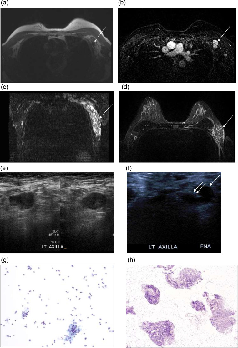
Axial T1-weighted sequence without fat saturation (A) and fat saturated, contrast enhanced MRI image (B) shows prominent left axillary lymph nodes with thickened cortex and absence of the fat in the hilum at level I (arrow). Bilateral T2-weighted short tau inversion recovery (STIR) reconstructed coronal (C) and axial (D) MRI demonstrate hyperintense T2 signal in the left axilla asymmetric compared to the right axilla, compatible with edema (arrow). Left axillary grayscale ultrasound, transverse plane (E,F) shows lymph node with thickened cortex, which underwent FNA (single arrow – lymph node; double arrow - needle). Magnified image of Pap-stained ThinPrep slide (G) showing polymorphous lymphoid population with follicular germinal center fragments. Magnified image of H&E-stained paraffin block (H) showing multiple fragments of benign lymphoid tissue with no evidence of metastatic carcinoma.
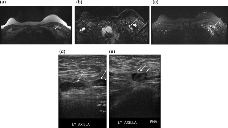
Axial T1-weighted sequence without fat saturation (A) and fat saturated, contrast enhanced MRI image (B) show a left axillary lymph node with lack of fat in the hilum, at level I (arrow). Bilateral T2-weighted short tau inversion recovery (STIR) MRI reconstructed axial (C) demonstrate hyperintense T2 signal in the left axilla asymmetric compared to the right axilla, compatible with edema. Bilateral axillary grayscale ultrasound, transverse plane (D,E) shows left-sided lymph node with thickened cortex but no edema is identified (D), which underwent FNA (single arrow – lymph node; double arrow - needle) (E). Magnified image of Pap-stained ThinPrep slide (F) showing polymorphous lymphoid population with follicular germinal center fragments.
References
-
- Newfield L, Naschitz JE, Yeshurun D. BCG-induced axillary lymph-adenitis in the adult. Harefuah. 1990;119(7-8):199–200. - PubMed
Publication types
LinkOut - more resources
Full Text Sources
