

Retinal Occlusive Vasculitis in a Patient with Hyperimmunoglobulin E Syndrome
- PMID: 34976422
- PMCID: PMC8718279
- DOI: 10.1155/2021/6317358
Retinal Occlusive Vasculitis in a Patient with Hyperimmunoglobulin E Syndrome
Abstract
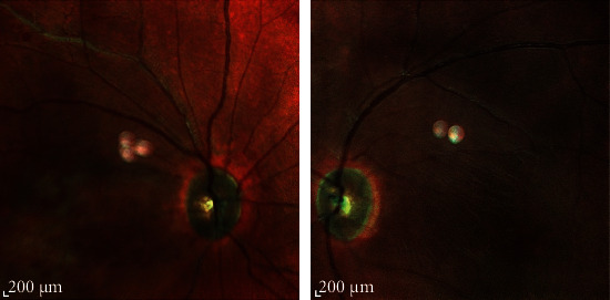
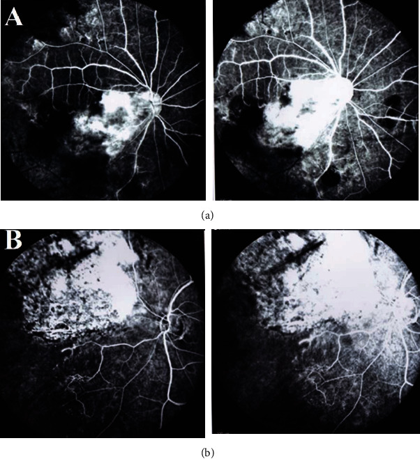
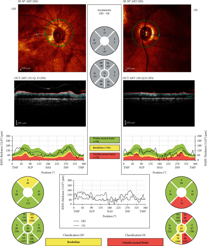
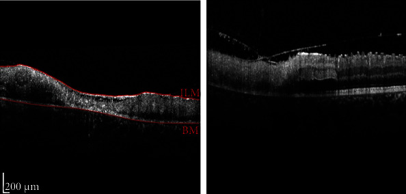
Background: Hyperimmunoglobulin E syndrome (HIES), or Job's syndrome, is a primary immunodeficiency disorder that is characterized by an elevated level of IgE with values reaching over 2000 IU (normal < 200 IU), eczema, and recurrent staphylococcus infection. Affected individuals are predisposed to infection, autoimmunity, and inflammation. Herein, we report a case of HIES with clinical findings of retinal occlusive vasculitis. Case Presentation. A 10-year-old boy with a known case of hyperimmunoglobulin E syndrome had exhibited loss of vision and bilateral dilated fixed pupil. Fundoscopic examination revealed peripheral retinal hemorrhaging, vascular sheathing around the retinal arteries and veins, and vascular occlusion in both eyes. A fluorescein angiography of the right eye showed hyper- and hypofluorescence in the macula and hypofluorescence in the periphery of the retina, peripheral arterial narrowing, and arterial occlusion. A fluorescein angiography of the left eye showed hyper- and hypofluorescence in the supranasal area of the optic disc. Macular optical coherence tomography of the right eye showed inner and outer retinal layer distortion. A genetic study was performed that confirmed mutations of the dedicator of cytokinesis 8 (DOCK 8). HSV polymerase chain reaction testing on aqueous humor and vitreous was negative, and finally, the patient was diagnosed with retinal occlusive vasculitis.
Conclusion: Occlusive retinal vasculitis should be considered as a differential diagnosis in patients with hyperimmunoglobulin E syndrome presenting with visual loss.
Copyright © 2021 Mohsen Farvardin et al.
Conflict of interest statement
The authors declare that they have no conflict of interest.
Figures
Similar articles
-
Retinal Vasculitis and Intraocular Inflammation after Intravitreal Injection of Brolucizumab.Ophthalmology. 2020 Oct;127(10):1345-1359. doi: 10.1016/j.ophtha.2020.04.017. Epub 2020 Apr 25. Ophthalmology. 2020. PMID: 32344075
-
Idiopathic Retinitis, Vasculitis, Aneurysms, and Neuroretinitis (IRVAN): Early Treatment Saves Sight.Cureus. 2022 Mar 10;14(3):e23049. doi: 10.7759/cureus.23049. eCollection 2022 Mar. Cureus. 2022. PMID: 35419227 Free PMC article.
-
Fungal infection of gingiva in a patient with hyperimmunoglobulin-E (Job's) syndrome.J Indian Soc Periodontol. 2012 Apr;16(2):256-60. doi: 10.4103/0972-124X.99272. J Indian Soc Periodontol. 2012. PMID: 23055595 Free PMC article.
-
MULTIPLE EVANESCENT WHITE DOT SYNDROME WITH CENTRAL VISUAL LOSS.Retin Cases Brief Rep. 2017 Winter;11 Suppl 1:S219-S225. doi: 10.1097/ICB.0000000000000467. Retin Cases Brief Rep. 2017. PMID: 27824724 Review.
-
RECURRENT BRANCH RETINAL ARTERY OCCLUSION FROM SUSAC SYNDROME: CASE REPORT AND REVIEW OF LITERATURE.Retin Cases Brief Rep. 2020 Fall;14(4):315-320. doi: 10.1097/ICB.0000000000000751. Retin Cases Brief Rep. 2020. PMID: 29870024 Review.
References
-
- Hsu A. P., Davis J., Puck J. M., Holland S. M., Freeman A. F. In: STAT3 Hyper IgE Syndrome . Adam M. P., Ardinger H. H., Pagon R. A., et al., editors. Seattle: Seattle (WA): University of Washington; 1993-2021.
-
- Engelhardt K. R., McGhee S., Winkler S., et al. Large deletions and point mutations involving the dedicator of cytokinesis 8 (DOCK8) in the autosomal-recessive form of hyper-IgE syndrome. The Journal of Allergy and Clinical Immunology . 2009;124(6):1289–1302.e4. doi: 10.1016/j.jaci.2009.10.038. - DOI - PMC - PubMed
-
- Hashemi H., Mohebbi M., Mehravaran S., Mazloumi M., Jahanbani-Ardakani H., Abtahi S.-H. Hyperimmunoglobulin E syndrome: genetics, immunopathogenesis, clinical findings, and treatment modalities. Journal of Research in Medical Sciences . 2017;22(1):p. 53. doi: 10.4103/jrms.jrms_1050_16. - DOI - PMC - PubMed
Publication types
LinkOut - more resources
Full Text Sources
