Review
. 2022 Jan 1;6(1):31-49.
doi: 10.7150/ntno.62970. eCollection 2022.

Advances in Surface Enhanced Raman Spectroscopy for in Vivo Imaging in Oncology

Affiliations
Review

Advances in Surface Enhanced Raman Spectroscopy for in Vivo Imaging in Oncology

Kenry et al. Nanotheranostics. .

Abstract

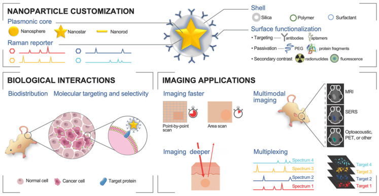
In the last two decades, the application of surface enhanced Raman scattering (SERS) nanoparticles for preclinical cancer imaging has attracted increasing attention. Raman imaging with SERS nanoparticles offers unparalleled sensitivity, providing a platform for molecular targeting, and granting multiplexed and multimodal imaging capabilities. Recent progress has been facilitated not only by the optimization of the SERS contrast agents themselves, but also by the developments in Raman imaging approaches and instrumentation. In this article, we review the principles of Raman scattering and SERS, present advances in Raman instrumentation specific to cancer imaging, and discuss the biological means of ensuring selective in vivo uptake of SERS contrast agents for targeted, multiplexed, and multimodal imaging applications. We offer our perspective on areas that must be addressed in order to facilitate the clinical translation of SERS contrast agents for in vivo imaging in oncology.


Conflict of interest statement

Competing Interests: The authors have declared that no competing interest exists.

Figures

Figure 1
Figure 1
Advances in SERS-based in vivo imaging. Top: The modular core-shell structure allows nanoparticle customization for strong SERS signal and functionalization for molecular targeting and multimodal contrast. Bottom left: Biological considerations improve nanoparticle pharmacodynamics and allow tumor selectivity and molecular targeting. Bottom right: New instrumentation and processes can lead to improved Raman imaging, as well as multimodal and multiplexed in vivo imaging.
Figure 2
Figure 2
Principles of Raman scattering, SERS, SERRS, and synthesis of a SERRS nanoparticle. (a) Jablonski diagram illustrating Raman scattering. (b) The Raman spectrum (“fingerprint”) of a compound has peaks corresponding to the chemical structure. (c) Gold nanostructures with their typical absorption spectra. (d) Examples of fluorescent dye absorption spectra. (e) Chemical structures of example chalcogenopyrylium-based Raman reporters (Dye 676, Dye 823, Dye 959 with optical absorbance at 676, 823, and 959 nm, respectively) and non-resonant reporters (BPE and AZPY). (f) Raman peak intensities of the reporters in (e) excited with an 830 nm laser source. (g) Schematic illustrating the different components of a SERRS nanoparticle and its synthesis process. Both the gold nanostar and the Raman reporter feature absorption maxima in the NIR. (e-f) Adapted with permission from ref. . Copyright 2018 The Royal Society of Chemistry.
Figure 3
Figure 3
Raman instrumentation. (a) Illumination pathway and collection pathway of the Small Animal Raman Imaging (SARI) instrument. The 785 nm laser excitation path is indicated by the red line and Raman scattered light by the yellow line. The SARI provides a 10-fold improvement in scan time compared with a traditional Raman microscope operating in the high-speed acquisition mode with matched spectral and spatial resolution. (b) In vivo SORS set up. A 785 nm laser was delivered at a 45˚ angle with regards to the collection optics. A translational xyz stage was used to move the laser away from the point of collection in order to apply the SORS technique. Detection of GBM in vivo through the intact skull is achieved using SESORS imaging as confirmed by MRI and ex vivo histology. (a) Reproduced with permission from ref. . Copyright 2013 United States National Academy of Sciences. (b) Reproduced with permission from ref. . Copyright 2019 Ivyspring International.
Figure 4
Figure 4
Untargeted SERRS Nanoparticles for Detection of Premalignant Lesions. (a-b) SERRS imaging of premalignant lesions in a KPC pancreatic cancer mouse model. (a) Photograph and the corresponding Raman images of the upper abdomen of a mouse with a pancreatic ductal adenocarcinoma (PDAC) in pancreas (top panels, outlined with a white dotted line) and excised pancreas (bottom panels) (b) H&E staining of the pancreas indicating PDAC and pancreatic intraepithelial neoplasia (PanIN) (arrows 1 and 2, respectively). Lesions in regions 1 and 2 were confirmed with histology and keratin 19 (KRT19) staining (c-g) SERRS imaging of premalignant lesions in an Apc gastrointestinal cancer mouse model. (c) Endoscopic images of polyp and normal tissue (labeled as “1” dashed line region and “2”, respectively) in the colon of a rat. (d) 2D Raman map of the SERRS signal intensity along the colon of the rat. (e) 3D projection of the 2D Raman map of the same colon of the rat. (f) White-light image of the evaluated colon ex vivo. (g) Histopathologic examination of the colon confirmed the correlation between positive SERRS signal and the presence of adenomatous polyps (labeled as “1” and “3”) as well as negative SERRS signal and the absence of lesions (labeled as “2”). Scale bars represent 2.5 mm. (a-b) Adapted with permission from ref. . Copyright 2015 American Association for the Advancement of Science. (c-g) Adapted with permission from ref. . Copyright 2019 American Chemical Society.
Figure 5
Figure 5
Topically applied SERRS ratiometry targeting the folate receptor. (a) Two distinct SERRS nanoparticles were synthesized: red - targeted against the folate receptor, blue - non targeted. (b) White light and bioluminescence imaging make the precise identification of the diffuse tumor difficult. (c) The decoupled signal from each of the two SERRS nanoparticles, does not offer any indication of the tumor site. (d) Ratiometry of the targeted probe over the untargeted, reveals the extend of the main tumor and multiple microtumors. Adapted with permission from ref. . Copyright 2017 American Chemical Society.
Figure 6
Figure 6
SERS nanoparticles developed for multimodal imaging. (a) A tri-modality MRI/Photoacoustic/Raman nanoparticle. The gold core provides photoacoustic contrast while Gadolinium, chelated on the surface, provides MRI contrast. (b) A PET/Raman nanoparticle. Gallium-68 embedded within the silica shell emits positrons allowing pre-operative whole-mouse imaging. The contrast comes from healthy liver tissue, with hypo-intense tumors. (c) A fluorescence/Raman nanoparticle based on a DNA linker for the fluorophore allows quick intraoperative tumor location with fluorescence, and highly precise margin definition with Raman imaging. (a) Adapted with permission from ref. . Copyright 2012 Nature Publishing Group. (b) Adapted with permission from ref. . Copyright 2017 Wiley-VCH. (c) Adapted with permission from ref. .

Similar articles

Cited by

References

    1. Andreou C, Kishore SA, Kircher MF. Surface-Enhanced Raman Spectroscopy: A New Modality for Cancer Imaging. Journal of Nuclear Medicine. 2015;56:1295. - PMC - PubMed
    1. Moriyasu F, Itoh K. Efficacy of perflubutane microbubble-enhanced ultrasound in the characterization and detection of focal liver lesions: phase 3 multicenter clinical trial. American Journal of Roentgenology. 2009;193:86–95. - PubMed
    1. Smeenge M, Tranquart F, Mannaerts CK, de Reijke TM, van de Vijver MJ, Laguna MP. et al. First-in-Human Ultrasound Molecular Imaging With a VEGFR2-Specific Ultrasound Molecular Contrast Agent (BR55) in Prostate Cancer: A Safety and Feasibility Pilot Study. Invest Radiol. 2017;52:419–27. - PubMed
    1. Carter LM, Poty S, Sharma SK, Lewis JS. Preclinical optimization of antibody-based radiopharmaceuticals for cancer imaging and radionuclide therapy—Model, vector, and radionuclide selection. Journal of Labelled Compounds and Radiopharmaceuticals. 2018;61:611–35. - PMC - PubMed
    1. Almuhaideb A, Papathanasiou N, Bomanji J. 18F-FDG PET/CT imaging in oncology. Ann Saudi Med. 2011;31:3–13. - PMC - PubMed

Publication types