. 2021 Dec 17:9:793073.
doi: 10.3389/fcell.2021.793073. eCollection 2021.

Characterizing the Neutrophilic Inflammation in Chronic Rhinosinusitis With Nasal Polyps

Affiliations

Characterizing the Neutrophilic Inflammation in Chronic Rhinosinusitis With Nasal Polyps

Jian-Wen Ruan et al. Front Cell Dev Biol. .

Erratum in

Abstract

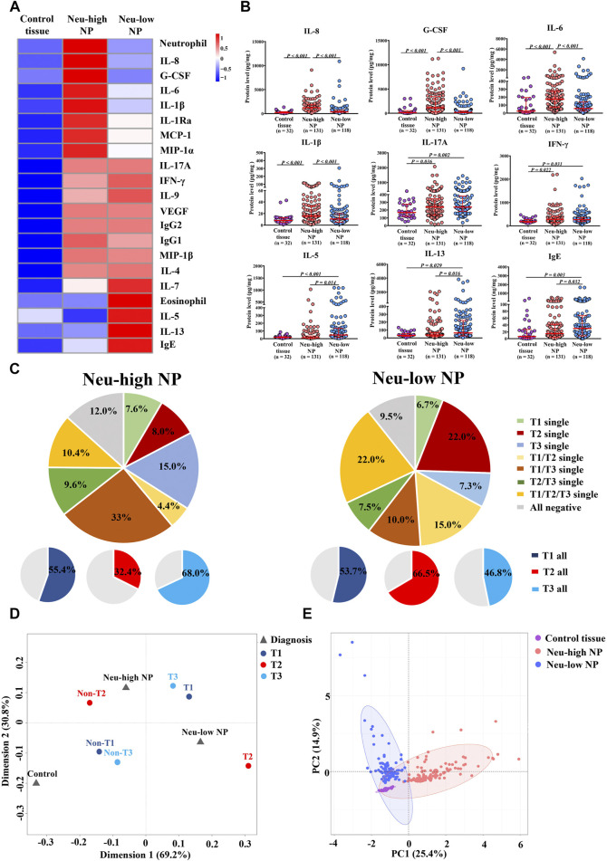
The mechanisms underlying neutrophilic inflammation in chronic rhinosinusitis with nasal polyps (CRSwNP) remain poorly investigated. This study aimed to examine the factors that contribute to tissue neutrophilia in CRSwNP. The numbers of neutrophils and active caspase-3-positive apoptotic neutrophils in sinonasal tissues were assessed via immunofluorescence staining. The 95th percentile of tissue neutrophil numbers in control subjects was selected as a cut-off to define neutrophil-high (Neu-high) or neutrophil-low (Neu-low) nasal polyps (NPs). The levels of 34 inflammatory mediators in sinonasal tissues were analyzed using Bio-Plex assay. Purified human peripheral blood neutrophils were incubated with nasal tissue homogenates, and the apoptotic neutrophils were assessed via flow cytometry. The cut-off for Neu-high NPs was >10 myeloperoxidase positive cells/high-power field. Compared with Neu-low NPs, Neu-high NPs had higher tissue levels of IL-1β, IL-1Ra, IL-6, IL-8, G-CSF, MCP-1, and MIP-1α, but lower levels of IL-5, IL-13, IgE, and eosinophils. Principal component and multiple correspondence analyses revealed mixed type 1, type 2, and type 3 endotypes for Neu-low NPs, and predominant type 1 and type 3 endotypes for Neu-high NPs. Neu-high NPs had lower percentages of apoptotic neutrophils than Neu-low NPs. The numbers of neutrophils and the percentages of apoptotic neutrophils correlated with G-CSF and IL-6 levels in the NPs. Tissue homogenates from Neu-high NPs, but not those from Neu-low NPs, suppressed neutrophil apoptosis in vitro, which was reversed by anti-G-CSF treatment. Tissue neutrophil numbers were associated with difficult-to-treat disease in patients with CRSwNP after surgery. We propose that G-CSF promotes neutrophilic inflammation by inhibiting neutrophil apoptosis in CRSwNP.

Keywords: apoptosis; chronic rhinosinusitis with nasal polyps; granulocyte colonystimulating factor; inflammation; neutrophil.


Conflict of interest statement

The authors declare that the research was conducted in the absence of any commercial or financial relationships that could be construed as a potential conflict of interest.

Figures

FIGURE 1
FIGURE 1
Distribution of tissue neutrophils in patients with CRSwNP. (A) Distribution of tissue neutrophils in control subjects (right panel) and CRSwNP patients (left panel). The red dotted lines represent 5th percentile (0.4) and 95th percentile (10.5) in the left panel. The red dotted lines represent minimum (0) and maximum (78.0), and the green dotted line represents the cut-off value (10) to define Neu-low and Neu-high NPs in the right panel. (B) Representative photomicrographs showing immunofluorescence staining of MPO positive cells and quantification of MPO positive cells. Original magnification ×400. Scale bar, 100 μm. Arrows denote representative positive cells. CRSwNP, chronic rhinosinusitis with nasal polyps; NP, nasal polyp; Neu-high, neutrophil-high; Neu-low, neutrophil-low; MPO, myeloperoxidase; HPF, high-power field.
FIGURE 2
FIGURE 2
Inflammatory features of Neu-high and Neu-low NPs. (A) Heatmap showing inflammatory mediators with different expression in at least one of the three groups as compared to other groups along with neutrophil and eosinophil numbers. (B) The levels of selected inflammatory mediators. (C) Patterns of T1, T2, and T3 endotype in Neu-high NPs (left panel) and Neu-low NPs (right panel). (D) Multiple correspondence analysis plot for the interrelationships between Neu-high NP, Neu-low NP, control tissue, and T1, T2 and T3 endotype. (E) Principal component analysis based on inflammatory mediators and cells indicated in heatmap. NP, nasal polyp; Neu-high, neutrophil-high; Neu-low, neutrophil-low; G-CSF, granulocyte colony-stimulating factor; IFN-γ, interferon γ; Ig, immunoglobulin; IL, interleukin; IL-1Ra, IL-1 receptor antagonist; MCP-1, monocyte chemoattractant protein-1; MIP, macrophage inflammatory protein; VEGF, vascular endothelial growth factor.
FIGURE 3
FIGURE 3
Correlations between tissue neutrophils, inflammatory cells, and mediators in the tissues and peripheral blood of patients with CRSwNP. A and B, Spearman correlation heatmap demonstrates unsupervised hierarchical clustering of the levels of tissue neutrophils and eosinophils, nine blood routine parameters, and 34 tissue inflammatory molecules in Neu-high (A) and Neu-low (B) NPs. Dendrograms are shown as trees, representing the distance between variables. The correlation matrix shows the positive (red) or negative (blue) correlation of two parameters. Color intensity reflects correlation strength. *p < 0.05, **p < 0.01, and ***p < 0.001. (C) Statistically significant correlations of tissue neutrophil numbers with the levels of cells and mediators in tissues of all patients with CRSwNP. NP, nasal polyp; Neu-high, neutrophil-high; Neu-low, neutrophil-low; bFGF, basic fibroblast growth factor; G-CSF, granulocyte colony-stimulating factor; IFN-γ, interferon γ; Ig, immunoglobulin; IL, interleukin; IL-1Ra, IL-1 receptor antagonist; IP-10, interferon-γ-induced protein-10; MCP-1, monocyte chemoattractant protein-1; MIP, macrophage inflammatory protein; PDGF-BB, platelet-derived growth factor-BB; TNF-α, tumor necrosis factor α; VEGF, vascular endothelial growth factor; HPF, high power field.
FIGURE 4
FIGURE 4
Reduced apoptosis of neutrophils in Neu-high NPs. (A) Representative immunofluorescence staining photomicrographs showing active caspase-3+MPO+ apoptotic neutrophils. Original magnification ×400. Arrows denote representative double positive cells. Scale bar, 100 μm. (B) The frequencies of active caspase-3+MPO+cells in total number of MPO+ cells. (C) The correlations between G-CSF (left panel) and lL-6 (right panel) levels and the percentages of active caspase-3+MPO+ cells in total NP tissues. NP, nasal polyp; Neu-high, neutrophil-high; Neu-low, neutrophil-low; MPO, myeloperoxidase; G-CSF, granulocyte colony-stimulating factor; IL-6, interleukin-6.
FIGURE 5
FIGURE 5
Neu-high NP environment inhibits neutrophil apoptosis. (A) Purified blood neutrophils (1 × 10⁶/well) from healthy subjects were cultured in the presence of homogenates (100 μg/ml) of control tissues, Neu-low NPs, and Neu-high NPs for 8 h and then subjected to flow cytometry. Representative flow cytometric analyses of Annexin-V+7-AAD- apoptotic, Annexin-V-7-AAD- live, and Annexin-V+7-AAD+ necrotic neutrophils are shown. (B) The mean frequencies of apoptotic, live, and necrotic neutrophils in different experimental groups are shown. (C–E) The frequencies of Annexin-V+7-AAD- apoptotic neutrophils (C), Annexin-V-7-AAD- live neutrophils (D), and Annexin-V+7-AAD+ necrotic neutrophils (E) after culture with homogenates of control tissues, Neu-high NPs, and Neu-low NPs. NP, nasal polyp; Neu-high, neutrophil-high; Neu-low, neutrophil-low; AAD, aminoactinomycin D.
FIGURE 6
FIGURE 6
G-CSF inhibits the apoptosis of neutrophils cultured with homogenates of Neu-high NPs. (A) Purified blood neutrophils (1 × 10⁶/well) from healthy subjects were cultured in the homogenates of Neu-high NPs in the presence of anti-G-CSF, anti-IL-6, or control IgG. Representative flow cytometric analyses of Annexin-V+7-AAD-apoptotic, Annexin-V-7-AAD- live, and Annexin-V+7-AAD+ necrotic neutrophils are shown. (B) The mean frequencies of apoptotic, live, and necrotic neutrophils in different experimental groups are shown. (C–E) The frequencies of Annexin-V+7-AAD- apoptotic neutrophils (C), Annexin-V-7-AAD- live neutrophils (D), and Annexin-V+7-AAD+ necrotic neutrophils (E) after culture with the homogenates of Neu-high NPs in the presence of anti-G-CSF, anti-IL-6 or control IgG. The data are analyzed in a paired fashion. G-CSF, granulocyte colony-stimulating factor; IL-6, interleukin-6; Ig, immunoglobulin; AAD, aminoactinomycin D; Neu-high, neutrophil-high.
FIGURE 7
FIGURE 7
Odds ratios for factors associated with difficult-to-treat CRSwNP. (A) Associations between difficult-to-treat disease and clinical features, and cellular and molecular markers, as analyzed via univariate regression analysis in patients with CRSwNP. (B) Associations between difficult-to-treat disease and clinical features, and cellular and molecular markers, as analyzed by multivariate regression analysis in patients with CRSwNP. The red frames delineate the neutrophil-associated factors. OR, odds ratio; CI, confidence interval; Ig, immunoglobulin; IL, interleukin; CRSwNP, chronic rhinosinusitis with nasal polyps.

References

    1. Arebro J., Drakskog C., Winqvist O., Bachert C., Kumlien Georén S., Cardell L. O. (2019). Subsetting Reveals CD16 High CD62L Dim Neutrophils in Chronic Rhinosinusitis with Nasal Polyps. Allergy 74 (12), 2499–2501. 10.1111/all.13919 - DOI - PubMed
    1. Bachert C., Zhang L., Gevaert P. (2015). Current and Future Treatment Options for Adult Chronic Rhinosinusitis: Focus on Nasal Polyposis. J. Allergy Clin. Immunol. 136 (6), 1431–1440. 10.1016/j.jaci.2015.10.010 - DOI - PubMed
    1. Bachert C., Zhang N., Holtappels G., De Lobel L., van Cauwenberge P., Liu S., et al. (2010). Presence of IL-5 Protein and IgE Antibodies to Staphylococcal Enterotoxins in Nasal Polyps Is Associated with Comorbid Asthma. J. Allergy Clin. Immunol. 126 (5), 962–968. 10.1016/j.jaci.2010.07.007 - DOI - PubMed
    1. Biffl W. L., Moore E. E., Moore F. A., Barnett C. C., Jr. (1995). Interleukin-6 Suppression of Neutrophil Apoptosis Is Neutrophil Concentration Dependent. J. Leukoc. Biol. 58 (5), 582–584. 10.1002/jlb.58.5.582 - DOI - PubMed
    1. Biondo C., Mancuso G., Midiri A., Signorino G., Domina M., Lanza Cariccio V., et al. (2014). The Interleukin-1β/CXCL1/2/Neutrophil Axis Mediates Host Protection against Group B Streptococcal Infection. Infect. Immun. 82 (11), 4508–4517. 10.1128/IAI.02104-14 - DOI - PMC - PubMed