Review
. 2021 Dec 15:8:799442.
doi: 10.3389/fsurg.2021.799442. eCollection 2021.

The Development of Laparoscopy-A Historical Overview

Affiliations
Review

The Development of Laparoscopy-A Historical Overview

Ibrahim Alkatout et al. Front Surg. .

Abstract

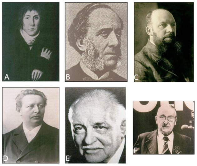
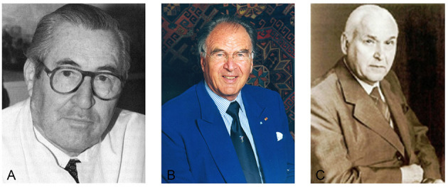
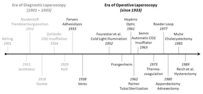
The advent of laparoscopy marked a fundamental change in the evolution of medicine. The procedure progressed consistently after the first time it was performed in a human being nearly a hundred years ago. The 1960's and 1980's witnessed groundbreaking changes. During this time, laparoscopy evolved from a purely diagnostic procedure into an independent surgical approach. Outstanding pioneers of the times were Palmer, Frangenheim and Semm. Laparoscopy advanced rapidly and influenced gynecology as well. The procedure was initially attacked most vociferously by the surgical fraternity. However, within a short period of time the pendulum shifted: laparoscopy became the preferred surgical approach for a variety of diseases-whether benign or malignant-in several medical disciplines. Laparoscopy has become a routine approach in the twenty-first century. Technical advancements have led to robot-assisted surgery. Future developments will include artificial intelligence and augmented reality. In the present article we address past milestones, current practices, and future challenges in laparoscopy.

Keywords: Kiel; Semm; history of medicine; kelling; laparoscopy; minimally invasive surgery.


Conflict of interest statement

The authors declare that the research was conducted in the absence of any commercial or financial relationships that could be construed as a potential conflict of interest. The handling editor RW declared a past co-authorship with one of the author IA.

Figures

Figure 1
Figure 1
Pioneers of laparoscopy. (A) Philipp Bozzini (1773–1809), (B) Antonin Jean Desormeaux (1815–1894), (C) Georg Kelling (1866–1945), (D) Maximilian Nitze (1848–1906), (E) Heinrich Kalk (1895–1973), (F) Raoul Palmer (1904–1985).
Figure 2
Figure 2
Pioneers of operative laparoscopy. (A) Hans Frangenheim (1920–2001), (B) Kurt Semm (1927–2003), (C) Karl Storz (1911–1996).
Figure 3
Figure 3
Introduction of intracorporeal knots (1974). Source: Department of Obstetrics and Gynecology, University Clinic of Kiel.
Figure 4
Figure 4
Timeline of the development of laparoscopy.
Figure 5
Figure 5
Pelvi trainer (1985). Source: Department of Obstetrics and Gynecology, University Clinic of Kiel.
Figure 6
Figure 6
Around 1970 Semm developed the “head ring,” a simple device by which he freed his left hand. The optical device need not be held; it hangs on a small hook. Source: Department of Obstetrics and Gynecology, University of Kiel.
Figure 7
Figure 7
Always with the finger on the pulse of the latest technology. As early as 1970, just a few months after the German tech company Philips launched their first “portable” TV camera, Semm used it in the operating room. Several educational films were produced with the aid of the camera (–43).
Figure 8
Figure 8
Technical breakthrough: electronic elements in the operating room at the Wertheim week in Kiel, organized by Semm in 1972. The operation was performed by Soichi Sakamoto and Semm and demonstrated by camera broadcast to a large number of national and international guests (camera work by Volker Rimkus, Semm's assistant). Source: Original interview with Volker Rimkus (born in 1939).
Figure 9
Figure 9
1972 Department of Obstetrics and Gynecology at the University Clinic of Kiel. The new technical options offered by the video camera enabled Semm to demonstrate his operations to a large number of national and international visiting doctors, who followed the operations with great interest. Semm referred to the event as the birth of video pelviscopy [33]. Source: Department of Obstetrics and Gynecology, University Clinic of Kiel.
Figure 10
Figure 10
Semm performing a sterilization by laparoscopy. Source: Department of Obstetrics and Gynecology, University of Kiel.
Figure 11
Figure 11
Laparoscopic appendectomy past and present. (A,B) are original pictures of the first laparoscopic appendectomy performed by Semm in 1980: skeletization of the appendix was followed by its ligation at the base with a Roeder loop. (C,D) show the current standard of surgery with the use of a stapler. The significantly better image quality and advanced instruments are evident. Source: Department of Obstetrics and Gynecology, University of Kiel.
Figure 12
Figure 12
Laparoscopic adnectomy past and present. (A,B) are original pictures of a laparoscopic adnectomy performed by Semm using the three-loop method in 1980. (C,D) show the current standard of surgery. The markedly better image quality is evident. Source: Department of Obstetrics and Gynecology, University of Kiel.
Figure 13
Figure 13
Original operation report by Semm in 1985. (A) Pelviscopy report of a patient who had undergone a hysterectomy, had chronic pain in the lower abdomen, suspected endometriosis of the external genitalia and extragenital endometriosis (endometriosis of the right-sided ovary, suspected endometriosis of the appendix). The patient underwent adhesiolysis, adnectomy on both sides by the 3-loop technique, and appendectomy. (B) Histological report showing evidence of a hemorrhagic corpus luteum cyst on the left side, endosalpingiosis of the left fallopian tube, and a scarred appendix. Source: Department of Obstetrics and Gynecology, University of Kiel.
Figure 14
Figure 14
International laparoscopy course at the Endoscopy School of Kiel with Professor Liselotte Mettler. Source: Department of Obstetrics and Gynecology, University of Kiel.
Figure 15
Figure 15
(A) Anatomical illustration of the uterus, adnexa and parametrium in an ancient anatomical atlas (Dr. Carl Toldt, Berlin and Wien 1903 [44]) and (B) modern female anatomical illustration generated by Markus Voll.

References

    1. Alkatout I, Holthaus B, Wedel T, Mettler L, Ackermann J, Maass N. Entwicklung der minimal-invasiven Chirurgie in der Gynäkologie und Überwindung assoziativer Herausforderungen. Der Gynäkologe. (2018) 51:737–43. 10.1007/s00129-018-4292-7 - DOI
    1. Alkatout I. An atraumatic retractor for interdisciplinary use in conventional laparoscopy and robotic surgery. Minim Invasive Ther Allied Technol. (2018) 27:265–71. 10.1080/13645706.2018.1440244 - DOI - PubMed
    1. Alkatout I, Mettler L. Hysterectomy a comprehensive surgical approach. J Turk Ger Gynecol Assoc. (2017) 18:221–3. 10.4274/jtgga.2017.0097 - DOI - PMC - PubMed
    1. Semm K. Pelviskopie und Hysteroskopie. Farbatlas und Lehrbuch. Stuttgart: F.K. Schattauer Verlag GmbH; (1976). p. 7–14.
    1. Schollmeyer T, Semm K, Schollmeyer M, Mettler L. Practical Manual for Laparoscopic Hysteroscopic Gynecological Surgery. 2 Edn. In: Schollmeyer T, Mettler L, Rüther D, et al.. eds. New Delhi: Jaypee Brothers Medical Publishers; (2013) p. 3–11. 10.5005/jp/books/11931_23 - DOI