. 2021 Dec 16;7(1):506-512.
doi: 10.1016/j.synbio.2021.12.003. eCollection 2022 Mar.

Fragment antigen binding domains (Fabs) as tools to study assembly-line polyketide synthases

Affiliations

Fragment antigen binding domains (Fabs) as tools to study assembly-line polyketide synthases

Katarina M Guzman et al. Synth Syst Biotechnol. .

Abstract

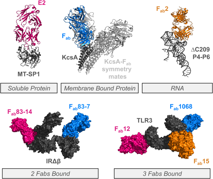
The crystallization of proteins remains a bottleneck in our fundamental understanding of their functions. Therefore, discovering tools that aid crystallization is crucial. In this review, the versatility of fragment-antigen binding domains (Fabs) as protein crystallization chaperones is discussed. Fabs have aided the crystallization of membrane-bound and soluble proteins as well as RNA. The ability to bind three Fabs onto a single protein target has demonstrated their potential for crystallization of challenging proteins. We describe a high-throughput workflow for identifying Fabs to aid the crystallization of a protein of interest (POI) by leveraging phage display technologies and differential scanning fluorimetry (DSF). This workflow has proven to be especially effective in our structural studies of assembly-line polyketide synthases (PKSs), which harbor flexible domains and assume transient conformations. PKSs are of interest to us due to their ability to synthesize an unusually broad range of medicinally relevant compounds. Despite years of research studying these megasynthases, their overall topology has remained elusive. One Fab in particular, 1B2, has successfully enabled X-ray crystallographic and single particle cryo-electron microscopic (cryoEM) analyses of multiple modules from distinct assembly-line PKSs. Its use has not only facilitated multidomain protein crystallization but has also enhanced particle quality via cryoEM, thereby enabling the visualization of intact PKS modules at near-atomic (3-5 Å) resolution. The identification of PKS-binding Fabs can be expected to continue playing a key role in furthering our knowledge of polyketide biosynthesis on assembly-line PKSs.

Keywords: Cryo-EM; Crystallography; Fragment antigen binding domains; Polyketide synthases.


Figures

Fig. 1
Fig. 1
Types of crystallization chaperones (CCs) for a protein of interest (POI).
Fig. 2
Fig. 2
Examples of structurally characterized Fab-bound proteins. Broad application of Fabs demonstrated: soluble (MT-SP1-E2; PDB ID 3BN9) [12] and membrane bound proteins (KcsA-Fab; PDB ID 1K4C) [13] and RNA (ΔC209 P4–P6-Fab2; PDB ID 2R8S) [14]. Use of multiple Fabs bound to one target protein. IRΔβ-Fab83-14-Fab83-7; PDB ID 4ZXB (left) [15]. TLR3-Fab1068-Fab12-Fab15; PDB ID 3ULV (right) [18]. In all cases: POI (gray); Fab (pink, orange, or blue).
Fig. 3
Fig. 3
High throughput methodology for determining advantageous Fabs for crystallography. Phage display libraries of Fabs are screened (ELISA) for potential binders to protein target (top), followed by DSF analysis of how the Fab alters the POI's physical properties (bottom).
Fig. 4
Fig. 4
Biosynthesis of 6-deoxyerythronolide B (6-dEB) by the 6-deoxyerythronolide B synthase (DEBS). DEBS is comprised of three large proteins, DEBS1, DEBS2 and DEBS3, each harboring two elongation modules. Each module performs one round of elongation and modification on the growing chain before translocating it to the next module. KS, ketosynthase; AT, acyltransferase; KR, ketoreductase; ACP, acyl carrier protein; DH, dehydratase; ER, enoyl reductase; TE, thioesterase. N- and C- terminal docking domains are shown in dark gray. KR3 is inactive.
Fig. 5
Fig. 5
Porcine mFAS (PDB ID 2VZ8) (top), “extended” architecture with dimeric KS/KS interface flanked by MAT domains on either end composing the elongation portion of the enzyme (gray). DH, ER, and KR domains outlined below in modification region (orange). DEBS DD(3)-KS3-AT3 (gray) bound to 1B2 (Fab, pink) (bottom). α,β represent the two subunits of the homodimer.

References

    1. Koide S. Engineering of recombinant crystallization chaperones. Curr Opin Struct Biol. 2009;19(4):449–457. doi: 10.1016/j.sbi.2009.04.008. - DOI - PMC - PubMed
    1. Air G.M., Webster R.G., Colman P.M., Laver W.G. Distribution of sequence differences in influenza N9 neuraminidase of tern and whale viruses and crystallization of the whale neuraminidase complexed with antibodies. Virology. 1987;160(2):346–354. doi: 10.1016/0042-6822(87)90005-5. - DOI - PubMed
    1. Ostermeier C., Iwata S., Ludwig B., Michel H. Fv fragment-mediated crystallization of the membrane protein bacterial cytochrome c oxidase. Nat Struct Biol. 1995;2(10):842–846. doi: 10.1038/nsb1095-842. - DOI - PubMed
    1. Hamers-Casterman C., Atarhouch T., Muyldermans S., Robinson G., Hamers C., Songa E.B., Bendahman N., Hamers R. Naturally occurring antibodies devoid of light chains. Nature. 1993;363(6428):446–448. doi: 10.1038/363446a0. - DOI - PubMed
    1. Barthelemy P.A., Raab H., Appleton B.A., Bond C.J., Wu P., Wiesmann C., Sidhu S.S. Comprehensive analysis of the factors contributing to the stability and solubility of autonomous human VH domains. J Biol Chem. 2008;283(6):3639–3654. doi: 10.1074/jbc.M708536200. - DOI - PubMed

LinkOut - more resources