. 2021 Dec 11;7(12):e08589.
doi: 10.1016/j.heliyon.2021.e08589. eCollection 2021 Dec.

High mobility group box-1 (HMGB-1) and its receptors in the pathogenesis of malaria-associated acute lung injury/acute respiratory distress syndrome in a mouse model

Affiliations

High mobility group box-1 (HMGB-1) and its receptors in the pathogenesis of malaria-associated acute lung injury/acute respiratory distress syndrome in a mouse model

Tachpon Techarang et al. Heliyon. .

Abstract

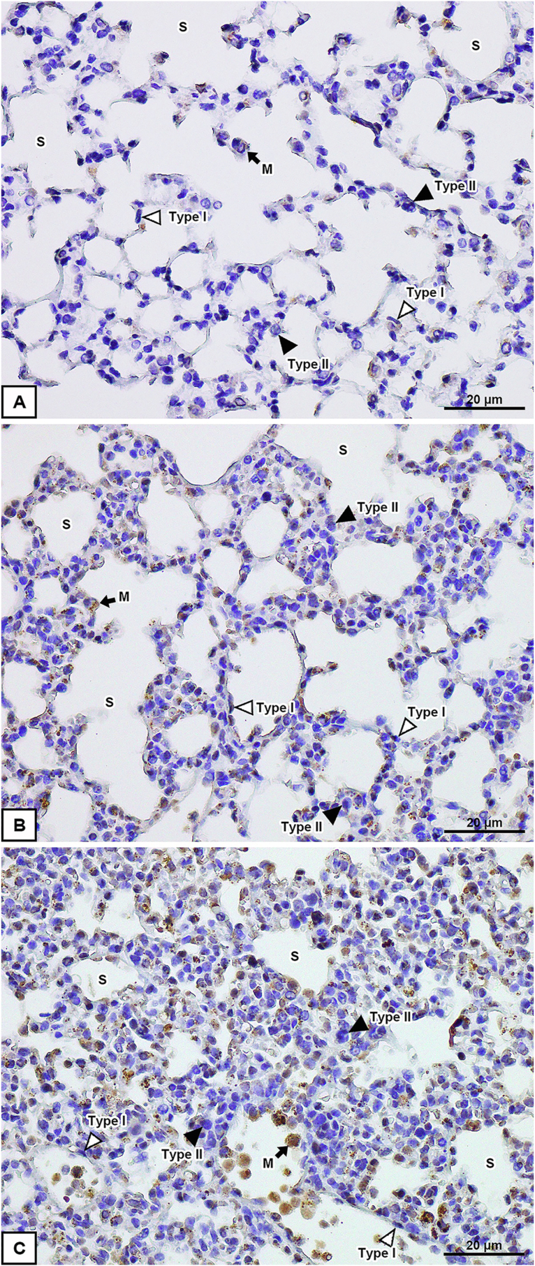
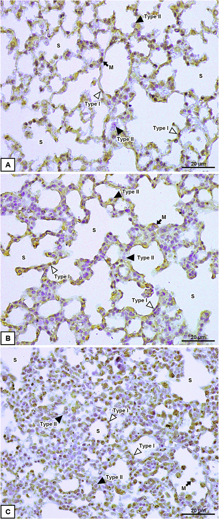
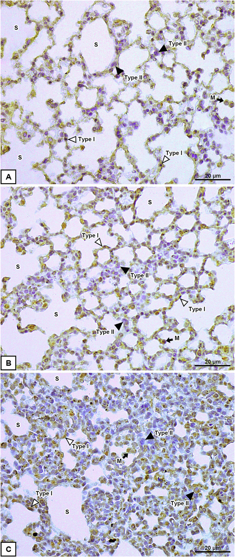
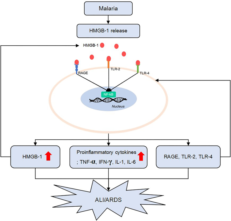
The DNA-binding protein high mobility group box-1 (HMGB-1) mediates proinflammatory cytokines that contribute to acute lung injury (ALI). Although ALI is a frequent complication of malaria infection, the contribution of HMGB-1 and its receptors to the pathogenesis of malaria-associated ALI/acute respiratory distress syndrome (MA-ALI/ARDS) has not been investigated in a mouse model. Here, the malaria-infected mice were divided into two groups according to lung injury score: the ALI/ARDS and non-ALI/ARDS groups. The expression of HMGB-1 and its receptors (RAGE, TLR-2 and TLR-4) in lung tissues was investigated by using immunohistochemical staining and real-time polymerase chain reaction (PCR). Additionally, HMGB-1 and proinflammatory cytokine (TNF-α, IFN-γ, IL-1 and IL-6) levels in plasma and lung tissues were quantified by using enzyme-linked immunosorbent assays. Cellular expression of both HMGB-1 and its receptors (RAGE, TLR-2 and TLR-4) was significantly increased in the lung tissues of the ALI/ARDS group compared with those in the non-ALI/ARDS and control groups. The levels of HMGB-1, TNF-α, IFN-γ, IL-1 and IL-6 were significantly increased in both plasma and lung tissues of the ALI/ARDS group compared with those in the non-ALI/ARDS and control groups, which were similar to the results obtained by real-time PCR. Increased mRNA expression of RAGE, TLR-2 and TLR-4 was found in the lung tissues of the ALI/ARDS group. Furthermore, the plasma HMGB-1 level was positively correlated with TLR-4 mRNA expression in the ALI/ARDS group. HMGB-1 levels were significantly increased in plasma and lung tissues of MA-ALI/ARDS mice and were related to the upregulated expression of HMGB-1 and proinflammatory cytokines. In conclusion, this study demonstrates that HMGB-1 is an important mediator of MA-ALI/ARDS pathogenesis and may represent a target for therapeutic malaria interventions with ALI/ARDS.

Keywords: Acute lung injury; Acute respiratory distress syndrome; HMGB-1; Malaria.


Conflict of interest statement

The authors declare no conflict of interest.

Figures

Figure 1
Figure 1
Histopathological changes in H&E-stained lung tissues in the control (A), malaria-infected without ALI/ARDS (B) and malaria-infected with ALI/ARDS (C and D) groups. Malaria-infected mice with ALI/ARDS showing alveolar oedema, thickening of alveolar septa and infiltration of leukocytes. Type I alveolar epithelial cell (Type I), Type II alveolar epithelial cell (Type II), alveolar macrophage (M), alveolar sac (S), capillary (C) and alveolar fibrin (F). All images were taken at 400× magnification. Bar = 20 μm.
Figure 2
Figure 2
Immunoperoxidase staining for HMGB-1 in lung tissues of the control (A), malaria-infected without ALI/ARDS (B) and malaria-infected with ALI/ARDS (C) groups. Type I alveolar epithelial cells (Type I), Type II alveolar epithelial cells (Type II), alveolar macrophages (M), and alveolar sacs (S). All images are 400× magnification. Bar = 20 μm.
Figure 3
Figure 3
Immunoperoxidase staining for RAGE in lung tissues of the control (A), malaria-infected without ALI/ARDS (B) and malaria-infected with ALI/ARDS (C) groups. Type I alveolar epithelial cells (Type I), Type II alveolar epithelial cells (Type II), alveolar macrophages (M), and alveolar sacs (S). All images are 400× magnification. Bar = 20 μm.
Figure 4
Figure 4
Immunoperoxidase staining for TLR-2 in lung tissues of the control (A), malaria-infected without ALI/ARDS (B) and malaria-infected with ALI/ARDS (C) groups. Type I alveolar epithelial cells (Type I), Type II alveolar epithelial cells (Type II), alveolar macrophages (M), and alveolar sacs (S). All images are 400× magnification. Bar = 20 μm.
Figure 5
Figure 5
Immunoperoxidase staining for TLR-4 in lung tissues of the control (A), malaria-infected without ALI/ARDS (B) and malaria-infected with ALI/ARDS (C) groups. Type I alveolar epithelial cells (Type I), Type II alveolar epithelial cells (Type II), alveolar macrophages (M), and alveolar sacs (S). All images are 400× magnification. Bar = 20 μm.
Figure 6
Figure 6
Levels of HMGB-1 and proinflammatory cytokines in the plasma and lung tissue of the control mice (n = 10), malaria-infected mice without ALI/ARDS (n = 17) and malaria-infected mice with ALI/ARDS (n = 11). The levels of HMGB-1 (A), IFN-γ (B), IL-1 (C), IL-6 (D), and TNF-α (E) were measured in plasma by ELISA. The levels of HMGB-1 (F), IFN-γ (G), IL-1 (H), IL-6 (I), and TNF-α (J) were measured in lung tissue by ELISA. ap < 0.001 compared with the control group. bp < 0.001 compared with the non-ALI/ARDS group. The data are presented as the mean ± SEM.
Figure 7
Figure 7
Schematic representation of HMGB-1 and its receptors in the pathogenesis of MA/ALIARDS.

References

    1. Lee S., Kwak M., Kim S., Shin J. The role of high mobility group box 1 in innate immunity. Yonsei Med. J. 2014;55:1165–1176. - PMC - PubMed
    1. Lotze M., Tracey K. High-mobility group box 1 protein (HMGB1): nuclear weapon in the immune arsenal. Nat. Rev. Immunol. 2005;5:331–342. - PubMed
    1. Lu B., Wang H., Andersson U., Tracey K.J. Regulation of HMGB1 release by inflammasomes. Protein Cell. 2013;4:163–167. - PMC - PubMed
    1. Andersson U., Tracey K.J. HMGB1 is a therapeutic target for sterile inflammation and infection. Annu. Rev. Immunol. 2011;29:139–162. - PMC - PubMed
    1. Higgins S.J., Xing K., Kim H., Kain D.C., Wang F., Dhabangi A., Musoke C., Cserti-Gazdewich C.M., Tracey K.J., Kain K.C., Liles W.C. Systemic release of high mobility group box 1 (HMGB1) protein is associated with severe and fatal Plasmodium falciparum malaria. Malar. J. 2013;12:105. - PMC - PubMed

LinkOut - more resources