. 2021 Oct 28;2(4):756-764.
doi: 10.1002/mco2.89. eCollection 2021 Dec.

Altering phosphoinositides in high-fat diet-associated prostate tumor xenograft growth

Affiliations

Altering phosphoinositides in high-fat diet-associated prostate tumor xenograft growth

Mingguo Huang et al. MedComm (2020). .

Abstract

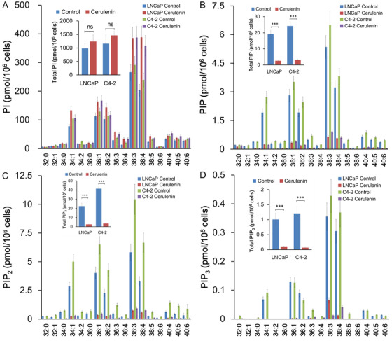
The metabolic reprogramming of phospholipids may affect intracellular signal transduction pathways. A high-fat diet (HFD) is attributed to prostate cancer (PCa) progression, but the expression pattern and role of phospholipids in HFD-mediated PCa progression remains unclear. In this study, HFD enhanced LNCaP xenograft tumor growth by upregulating the phosphatidylinositol (PI) 3-kinase (PI3K)/AKT signaling pathway. A lipidomic analysis using xenograft tumors showed that phosphoinositides, especially PI (3,4,5)-trisphosphate (PIP3), including several species containing C38:4, C38:3, and C40:4 fatty acids, increased in the HFD group compared to control. Fatty acid synthase (FASN) was significantly upregulated in xenograft tumors under HFD in both gene and protein levels. PCa cell growth was significantly inhibited through the decreased AKT signaling pathway by treatment with cerulenin, a chemical FASN inhibitor, which also downregulated PIP, PIP2, and PIP3 but not PI. Thus, dietary fat influences PCa progression and alters phosphoinositides, especially PIP3, a critical player in the PI3K/AKT pathway. These results may offer appropriate targets, such as FASN, for dietary intervention and/or chemoprevention to reduce PCa incidence and progression.

Keywords: AKT; FASN; high‐fat diet; phosphoinositide; prostate cancer.


Conflict of interest statement

The authors declare that they have no conflict of interest.

Figures

FIGURE 1
FIGURE 1
High‐fat diet (HFD) enhanced tumor growth in LNCaP xenograft mice with the upregulated PI3K/AKT pathway. LNCaP xenograft mice were subcutaneously injected with tumor cells and fed with HFD or control diet. On week 14, mice were sacrificed, and xenograft tumors were excised to performed lipid analysis. (A) The tumor volume level was enhanced in the HFD group compared to control. (B) Xenograft tumor sections were subjected to anti‐Ki‐67 staining. Ki‐67 positivity was significantly higher in the HFD group than the control group. Bar, 100 μm. (C) The serum insulin level, as measured by the specific ELISA kit, was no statistical difference in the diet groups. (D) Mouse blood glucose levels were quantified using LAB Gluco. There was no significant difference in blood glucose levels between the diet groups. (E) Xenograft tumor sections from mice in the two diet groups underwent immunohistological staining with anti‐human pAKT (Ser473) and pan‐AKT antibodies. The expression was semiquantitatively calculated. pAKT staining intensities were significantly higher in the HFD group than the control group. Bar, 100 μm. (F) pAKT (Ser473 and Thr308), AKT, and FASN expression in lysates of individual LNCaP xenograft tumors (three samples each group) was detected by Western blot using specific antibodies. β‐actin was used as an intrinsic control
FIGURE 2
FIGURE 2
High‐fat diet (HFD) affected phosphoinositide metabolism in LNCaP xenograft tissue. (A–D) Total lipids from mouse xenograft tumor tissue (10 mg; seven mice per group) were extracted. Total PI (A), PIP (B), PIP2 (C), and PIP3 (D) were quantified by liquid chromatography‐mass spectrometry (LC‐MS)/MS analysis. Abbreviation: ns, not significant.
FIGURE 3
FIGURE 3
Fatty acid synthase (FASN) was upregulated in LNCaP xenograft tumors under HFD feeding and regulated cell growth and pAKT expression in PCa cells. The expression of 84 key genes involved in regulating fatty acid metabolism was comprehensively analyzed by a Human Fatty Acid Metabolism PCR Array kit using xenograft tumors. (A) The cluster heat map shows the relative gene expression between the diet groups. The assay was performed in duplicate within each group. Wells a1 to g12 each contain real‐time PCR array for the fatty acid metabolism‐related gene. Increased levels are red, and decreased levels are green. (B) FASN was significantly upregulated in xenograft tumors under HFD feeding. (C and D) Role of FASN in PCa cell viability and AKT expression. LNCaP and C4‐2 cells were cultured with or without 10 μM cerulenin for 24 h. (C) Cell proliferation was assessed using a nonradioactive MTT‐based Cell Proliferation Assay kit, and cell viability in cells treated with cerulenin was compared to untreated cells. *< 0.05. (D) pAKT (Ser473 and Thr308) and AKT expression in lysates (10 μg) were detected by Western blot using specific antibodies. β‐actin was used as an intrinsic control. ***< 0.001
FIGURE 4
FIGURE 4
FASN regulated phosphoinositides in PCa cells. (A–D) LNCaP and C4‐2 cells (1 × 10⁶) were seeded in a 60‐mm dish and treated with 10 μM cerulenin for 24 h. Total lipids from cultured cells were extracted, and PI and PIPs were quantified. The expression levels of PIP (B), PIP2 (C), and PIP3 (D), but not PI (A), were decreased in PCa cells treated with cerulenin compared to untreated cells. ***< 0.001
FIGURE 5
FIGURE 5
Orlistat decreased PIPs levels but not PI in PCa C4‐2 cells. (A–D) C4‐2 cells (1 × 10⁶) were seeded in a 60 mm dish and treated with 10 μM orlistat for 24 h. Total lipids from cultured cells were extracted, and PI and PIPs were quantified. The expression levels of PIP (B), PIP2 (C), and PIP3 (D), but not PI (A), were decreased in PCa cells treated with orlistat compared to untreated cells. (E) Schematic of the putative effect of HFD and/or obesity on the PCa progression. HFD‐induced alteration of PIPs, especially PIP3, may activate proliferative signaling transduction, such as the PI3K/AKT pathway through the upregulation of FASN, and influence PCa progression. Thus, PI3K/AKT signaling and FASN could be the important targets for dietary intervention and/or chemoprevention in PCa incidence and progression

References

    1. Jemal A, Siegel R, Ward E, Murray T, Xu J, Thun MJ. Cancer statistics. CA Cancer J Clin. 2007;57(1):43‐66. - PubMed
    1. Ito K. Prostate cancer in Asian men. Nat Rev Urol. 2014;11(4):197‐212. - PubMed
    1. Kolonel LN. Fat, meat, and prostate cancer. Epidemiol Rev. 2001;23(1):72‐81. - PubMed
    1. Eisinger K, Krautbauer S, Hebel T, et al. Lipidomic analysis of the liver from high‐fat diet induced obese mice identifies changes in multiple lipid classes. Exp Mol Pathol. 2014;97(1):37‐43. - PubMed
    1. Narita S, Tsuchiya N, Saito M, et al. Candidate genes involved in enhanced growth of human prostate cancer under high fat feeding identified by microarray analysis. Prostate. 2008;68(3):321‐335. - PubMed