Review
. 2021:1:100005.
doi: 10.1016/j.hsr.2021.100005. Epub 2021 Dec 3.

Mucormycosis: A triple burden in patients with diabetes during COVID-19 Pandemic

Affiliations
Review

Mucormycosis: A triple burden in patients with diabetes during COVID-19 Pandemic

A S Fathima et al. Health Sci Rev (Oxf). 2021.

Abstract

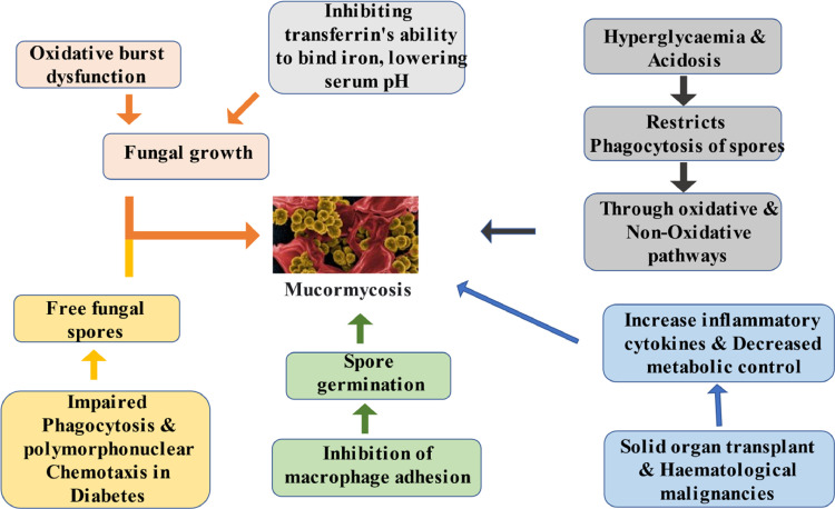
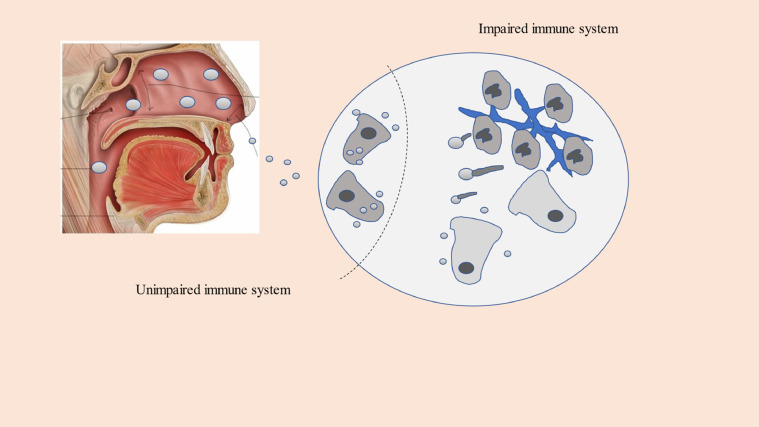
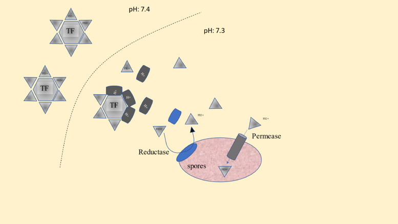
With the upsurge in the cases of COVID-19 around the world, plenty of potential COVID-19 complications are becoming more prevalent, including a higher risk of secondary bacterial and fungal infections. Mucormycosis is one such condition which has high prevalence among individuals with diabetes who were infected with COVID-19.The usage of steroids in the treatment further inflates the risk of infection and exacerbation of disease in pre-existent mucormycosis patients. Generally, Corticosteroid-induced diabetes can arise on long-term steroid medication, increasing the likelihood of mucormycosis. In patients with COVID-19, the indications and dose of corticosteroids should be properly regulated, and persons with diabetes who take insulin or oral anti-diabetic medicines should be cautious. To avoid poor outcomes, strategies to improve glycemic management should be emphasized. This narrative review elucidates different disciplines on rampant use of steroids, iron and zinc supplements as well as the methods utilized as primary or adjunctive treatment of this fatal condition. This article may help to pave the way for robust research that needs to be done to tackle the deadly triple burden of the disease.

Keywords: COVID-19; Diabetes; Mucormycosis; Triple burden.


Conflict of interest statement

The authors declare that they have no known competing financial interests or personal relationships that could have appeared to influence the work reported in this paper.

Figures

Fig. 1
Fig. 1
Major causative pathways of Mucormycosis in persons with diabetes.
Fig. 2
Fig. 2
Excess ferritin levels leading to fungal growth and Mucormycosis.
Fig. 3
Fig. 3
Impaired immune system Figure 2 The impaired cellular pathways responsible for Mucorales invasion in diabetes. A. Spores enter the organism by inhalation, cutaneous inoculation or ingestion. B. In cases of a competent immune system, macrophages mediated by the alternative complement pathway (black triangles) engulf spores and prevent their germination. C. Phagocytosis and polymorphonuclear (PMN) chemotaxis are impaired in diabetes, and macrophages are unable to phagocytose spores, resulting in free spores (1) in tissues. (2) The spores swell and form buds (germ-tube growth; 3) which elongate into hyphae (4). PMN are recruited, however oxidative burst dysfunction is linked to fungal growth, vascular invasion, and eventual haematogenous dissemination. (Adapted from Rammaert et al.) .
Fig. 4
Fig. 4
Role of iron in pathogenesis of Mucormycosis Fig. 4: The pathogenesis of Mucormycosis is connected to changes in iron metabolism in diabetes. The binding protein transferrin transports ferric iron (Fe3+) (TF). TF iron-binding capacity is reduced by proton (H+) efflux in low pH situations, such as ketoacidosis, and free iron is released into the bloodstream (explained in Fig. 4).This accessible iron is reduced to ferrous iron (Fe2+) by a Mucorales spore membrane reductase enzyme, which solubilizes free iron to allow it to penetrate spores through the high-affinity permease FTR1, resulting in a ferric form that aids the growth of Mucorales spore .

References

    1. Ibrahim A.S., Spellberg B., Walsh T.J. Kontoyiannis DP. Pathogenesis of mucormycosis. Clin. Infect. Dis. 2012;54(suppl_1):S16–S22. doi: 10.1093/cid/cir865. Feb 1. - DOI - PMC - PubMed
    1. Herrera D.A., Dublin A.B., Ormsby E.L., Aminpour S., Howell L.P. Imaging findings of rhinocerebral mucormycosis. Skull Base. 2009;19(02):117–125. doi: 10.1055/s-0028-1096209. Apr. - DOI - PMC - PubMed
    1. Moorthy A., Gaikwad R., Krishna S., Hegde R., Tripathi K.K., Kale P.G., Rao P.S., Haldipur D., Bonanthaya K. SARS-CoV-2, uncontrolled diabetes and corticosteroids—an unholy trinity in invasive fungal infections of the maxillofacial region? A retrospective, multi-centric analysis. J. Maxillofac. Oral Surg. 2021:1–8. doi: 10.1007/s12663-021-01532-1. Mar 6. - DOI - PMC - PubMed
    1. Ravani S.A., Agrawal G.A., Leuva P.A., Modi P.H., Amin K.D. Rise of the phoenix: mucormycosis in COVID-19 times. Indian J. Ophthalmol. 2021;69(6):1563–1568. doi: 10.4103/ijo.IJO_310_21. Jun 1. - DOI - PMC - PubMed
    1. Petrikkos George, Skiada Anna, Lortholary Olivier, Roilides Emmanuel, Walsh Thomas J., Kontoyiannis Dimitrios P. Epidemiology and clinical manifestations of mucormycosis. Clin. Infect. Dis. 2012;54(1):S23–S34. doi: 10.1093/cid/cir866. - DOI - PubMed