

Does new bone formation vary in different sites within the same maxillary sinus after lateral augmentation? A prospective histomorphometric study
- PMID: 34978096
- PMCID: PMC9306466
- DOI: 10.1111/clr.13891
Does new bone formation vary in different sites within the same maxillary sinus after lateral augmentation? A prospective histomorphometric study
Abstract
Objective: The aim of this study was to evaluate histomorphometric outcomes of lateral maxillary sinus augmentation in different areas of the same cavity and to correlate results to bucco-palatal sinus width (SW) and residual bone height (RBH).
Material and methods: Patients needing maxillary sinus floor elevation (RBH <5 mm) to insert two nonadjacent implants were treated with lateral augmentation using a composite graft. Six months later, two bone-core biopsies (mesial/distal) were retrieved in implant insertion sites. SW and RBH were measured on cone beam computed tomography, and correlations between histomorphometric and anatomical parameters were evaluated by multivariate linear regression analysis.
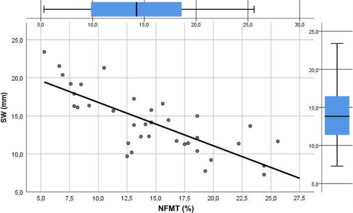
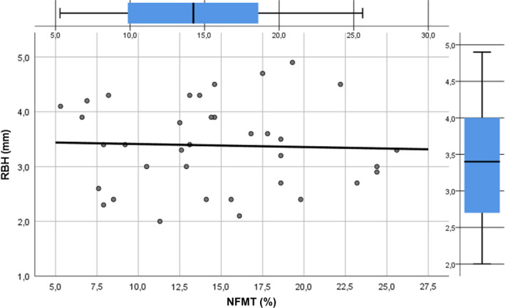
Results: Twenty patients underwent sinus augmentation, and eighteen were included in the final analysis (two dropouts for membrane perforation). Mean newly formed mineralized tissue percentage (%NFMT) after 6 months in mesial and distal sites was 17.5 ± 4.7 and 11.6 ± 4.7, respectively (p = .0004). Multivariate linear regression showed a strong negative correlation between SW and %NFMT (β coefficient=-.774, p < .0001) and no correlation between RBH and %NFMT (β coefficient =-.038, p = .825).
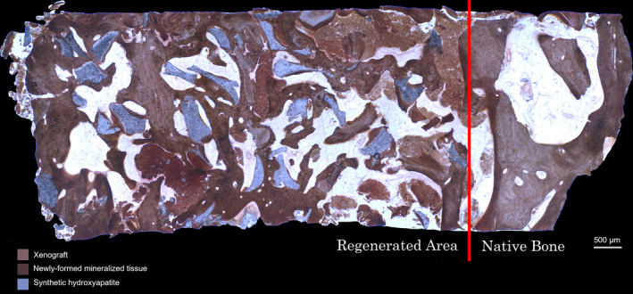
Conclusions: The present study confirms that %NFMT after lateral sinus augmentation occurs at different rates in different anatomical areas of the same maxillary sinus, showing a strong negative correlation with SW, whereas no influence of RBH was observed. Clinicians should regard SW as a guide for graft selection and to decide duration of the healing period. Researchers should consider SW as a predictor variable, when comparing regenerative outcomes of different biomaterials by using maxillary sinus as an experimental model.
Keywords: bone regeneration; bone substitutes; guided tissue regeneration; maxillary sinus; sinus floor elevation.
© 2022 The Authors. Clinical Oral Implants Research published by John Wiley & Sons Ltd.
Conflict of interest statement
The authors do not have any financial interests, either directly or indirectly, in the products or information listed in this paper.
Figures
Similar articles
-
New bone formation after transcrestal sinus floor elevation was influenced by sinus cavity dimensions: A prospective histologic and histomorphometric study.Clin Oral Implants Res. 2018 May;29(5):465-479. doi: 10.1111/clr.13144. Epub 2018 Mar 23. Clin Oral Implants Res. 2018. PMID: 29569763
-
Influence of Maxillary Sinus Width on New Bone Formation After Transcrestal Sinus Floor Elevation: A Proof-of-Concept Prospective Cohort Study.Implant Dent. 2017 Apr;26(2):209-216. doi: 10.1097/ID.0000000000000554. Implant Dent. 2017. PMID: 28125520
-
The effect of anatomy on osteogenesis after maxillary sinus floor augmentation: a radiographic and histological analysis.Clin Oral Investig. 2021 Sep;25(9):5197-5204. doi: 10.1007/s00784-021-03827-6. Epub 2021 Feb 11. Clin Oral Investig. 2021. PMID: 33569678
-
An overview on bone reconstruction of atrophic maxilla: success parameters and critical issues.J Biol Regul Homeost Agents. 2016 Apr-Jun;30(2 Suppl 1):209-15. J Biol Regul Homeost Agents. 2016. PMID: 27469570 Review.
-
Residual Bone Height and New Bone Formation after Maxillary Sinus Augmentation Procedure Using Biomaterials: A Network Meta-Analysis of Clinical Trials.Materials (Basel). 2023 Feb 6;16(4):1376. doi: 10.3390/ma16041376. Materials (Basel). 2023. PMID: 36837005 Free PMC article. Review.
Cited by
-
Clinical and radiographic outcomes following transcrestal maxillary sinus floor elevation with injectable xenogenous bone substitute in gel form: a prospective multicenter study.Int J Implant Dent. 2022 Jul 22;8(1):32. doi: 10.1186/s40729-022-00431-5. Int J Implant Dent. 2022. PMID: 35867239 Free PMC article.
-
Intraoperative complications and early implant failure after transcrestal sinus floor elevation with residual bone height ≤5 mm: A retrospective multicenter study.Clin Oral Implants Res. 2022 Aug;33(8):783-791. doi: 10.1111/clr.13959. Epub 2022 May 29. Clin Oral Implants Res. 2022. PMID: 35578774 Free PMC article.
-
Minimally invasive techniques for lateral maxillary sinus floor elevation: small lateral window and one-stage surgery-a 2-5-year retrospective study.Int J Oral Sci. 2023 Jul 11;15(1):28. doi: 10.1038/s41368-023-00233-4. Int J Oral Sci. 2023. PMID: 37433766 Free PMC article.
-
Bone Graft Packing and Its Association with Bone Regeneration in Maxillary Sinus Floor Augmentations: Histomorphometric Analysis of Human Biopsies.Biology (Basel). 2022 Sep 30;11(10):1431. doi: 10.3390/biology11101431. Biology (Basel). 2022. PMID: 36290335 Free PMC article.
-
Finite Element Analysis of Functionally Loaded Subperiosteal Implants Evaluated on a Realistic Model Reproducing Severe Atrophic Jaws.Methods Protoc. 2025 Jan 18;8(1):8. doi: 10.3390/mps8010008. Methods Protoc. 2025. PMID: 39846694 Free PMC article.
References
-
- Artzi, Z. , Kozlovsky, A. , Nemcovsky, C. E. , & Weinreb, M. (2005). The amount of newly formed bone in sinus grafting procedures depends on tissue depth as well as the type and residual amount of the grafted material. Journal of Clinical Periodontology, 32, 193–199. 10.1111/j.1600-051X.2005.00656.x - DOI - PubMed
-
- Avila, G. , Wang, H. L. , Galindo‐Moreno, P. , Misch, C. E. , Bagramian, R. A. , Rudek, I. , Benavides, E. , Moreno‐Riestra, I. , Braun, T. , & Neiva, R. (2010). The influence of the bucco‐palatal distance on sinus augmentation outcomes. Journal of Periodontology, 81, 1041–1060. - PubMed
-
- Avila‐Ortiz, G. , Wang, H. L. , Galindo‐Moreno, P. , Misch, C. E. , Rudek, I. , & Neiva, R. (2012). Influence of lateral window dimensions on vital bone formation following maxillary sinus augmentation. International Journal of Oral and Maxillofacial Implants, 27, 1230–1238. - PubMed
MeSH terms
LinkOut - more resources
Full Text Sources
