. 2022 Jan 3;5(1):2.
doi: 10.1186/s42155-021-00269-9.

Management of Pancreatico-duodenal arterio-venous malformation

Affiliations

Management of Pancreatico-duodenal arterio-venous malformation

Clement Marcelin et al. CVIR Endovasc. .

Abstract

Purpose: To describe the interventional management and clinical outcome of pancreatico-duodenal arterio-venous malformations (PDAVMs).

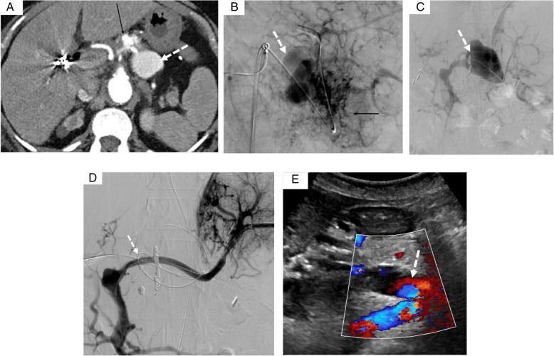
Material and methods: Seven patients presenting a PDAVM (6 women, 1 male; mean age: 61) were retrospectively reviewed. Technical, clinical success and complications of embolization and surgical management of symptomatic PDAVMs were assessed. Technical success was defined as a complete occlusion of the PDAVM and clinical success as no clinical symptom or recurrence during follow-up. Patients with asymptomatic PDAVMs were followed clinically, by Doppler ultrasound and CT-angiography.

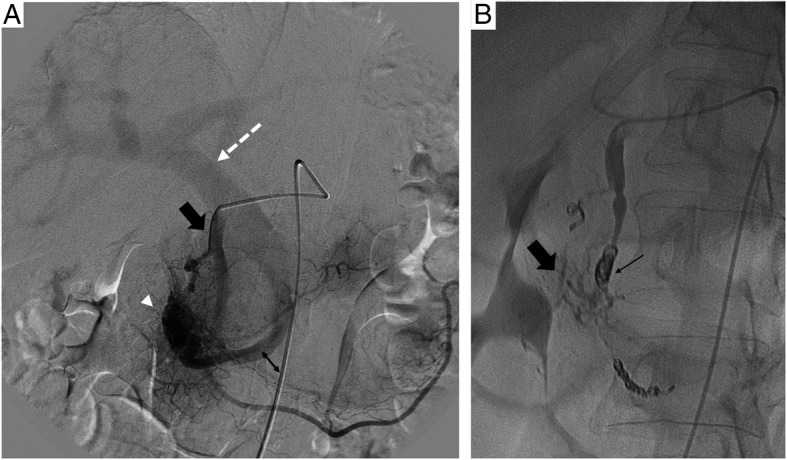
Results: Mean follow-up time was 69 months (15-180). Five symptomatic patients presented with upper gastrointestinal bleeding (n=3), ascites (n=1), and abdominal pain (n=1). Two patients were asymptomatic. The PDAVMs were classified as follow: Yakes I (1), IIIa (2), IIIb (3) and IV (1). Five symptomatic patients were treated with 9 embolization sessions with arterial approach (onyx®, glue, coils) in 7 and venous approach in 2 (plugs, coils, covered stents, STS foam and onyx®). Technical success of embolization was 60% (3/5). Devascularization was incomplete for 2 Yakes IIIB patients. Clinical success of embolization was estimated at 80% (4/5) as one patient required additional surgery (Whipple) because of persistent bleeding. One splenic vein thrombosis was treated successfully by mechanical thrombectomy and heparin. No recurrence occurred during follow-up. No progression was documented in asymptomatic patients.

Conclusion: Embolization of symptomatic PDAVMs is effective and surgery should be performed in second intention. Complete devascularization is more difficult to obtain in Yakes III PDAVM.

Keywords: AVM; Embolization; Pancreas; Percutaneous.


Figures

Fig. 1
Fig. 1
A 79 yo women with upper GI bleeding. A- Arteriography showed a type IIIa pancreatic AVM, vascularized by the gastro-duodenal artery (black arrow), connected to the gastro-duodenal vein (double arrow) with a nidus (arrowhead), draining into the portal vein (dotted arrow). B- After embolization of the gastroduodenal artery with coils (black arrow) and the nidus with onyx (large black arrow), opacification of the celiac trunk showed no residual vascularization of the AVM.
Fig. 2
Fig. 2
A 57 yo women with upper GI bleeding. A- CT scan showed a nidus in the pancreatic head (dotted arrow). B- Selective angiography of the pancreatic dorsal artery (black arrow) showing a type IIIB pancreatic AVM, with multiple feeding arteries draining in an aneurysmal vein (white arrow) both draining into the portal vein. C- Selective angiography of the gastroduodenal artery showing multiple arterial collaterals (black arrow), vascularized by multiples branches of the postero-superior and antero-superior pancreatico-duodenal arteries (white arrow). Embolization with Onyx® of the dorsal pancreatic artery (large black arrow). D- Portal venous access showed an enlarged pancreatic vein (black arrow) draining into the portal vein (big black arrow). E- Pressure cooker technique: proximal embolization of the draining gastroduodenal vein using a plug (white arrow), and then distal embolization with STS using a microcatheter distal to the Plug in order to reflux into the nidus (black arrow) of the AVM. F- Selective angiography of the celiac trunk showing a residual pancreatic AVM (arrow).
Fig. 3
Fig. 3
A 66 yo women with a cryptogenic cirrhosis who underwent previous abdominal surgery for colorectal carcinoma and chronic portal vein thrombosis presented recurrent ascites and chronic pancreatitis. A CT scans showing a Yakes type IIIa pancreatic AVM (arrow), with an aneurysmal splenic vein (dashed arrow). B Selective angiography of the splenic artery showed a pancreatic AVM, vascularized by the dorsal pancreatic artery (arrow), splenic artery, left gastric artery, and connected to an aneurysmal splenic vein (dashed arrow). C After puncture of splenic vein, venography showing the aneurysmal splenic vein draining into the gastroduodenal and mesenteric veins because of the preexisting portal thrombosis (dashed arrow). D Insertion of a covered stent in the splenic vein (dashed arrow) by a transplenic access and embolization using Onyx® and coils after direct puncture of the aneurysm. The patient had subsequent splenic venous thrombosis which was successfully treated by mechanical thrombectomy and heparin infusion. E Doppler ultrasound at 1 year showed permeability of the splenic and portal veins (dashed arrow), with no residual AVM.
Fig. 4
Fig. 4
Yakes AVM classification (Soulez et al., 2019)

References

    1. Aina R, Oliva VL, Therasse É, Perreault P, Bui BT, Dufresne M-P, et al. Arterial Embolotherapy for Upper Gastrointestinal Hemorrhage: Outcome Assessment. J Vasc Interv Radiol. 2001;12(2):195–200. doi: 10.1016/S1051-0443(07)61825-9. - DOI - PubMed
    1. Beyer LP, Wohlgemuth WA, Uller W, Pregler B, Goessmann H, Niessen C, et al. Percutaneous treatment of symptomatic superior mesenteric vein stenosis using self-expanding nitinol stents. Eur J Radiol. 2015;84(10):1964–1969. doi: 10.1016/j.ejrad.2015.06.013. - DOI - PubMed
    1. Cassinotto C, Lapuyade B. Pancreatic Arteriovenous Malformation Embolization with Onyx. J Vasc Interv Radiol. 2015;26(3):442–444. doi: 10.1016/j.jvir.2014.11.036. - DOI - PubMed
    1. Chapot R, Stracke P, Velasco A, Nordmeyer H, Heddier M, Stauder M, et al. The Pressure Cooker Technique for the treatment of brain AVMs. J Neuroradiol. 2014;41(1):87–91. doi: 10.1016/j.neurad.2013.10.001. - DOI - PubMed
    1. Cho SK, Do YS, Shin SW, Kim D-I, Kim YW, Park KB, et al. Arteriovenous Malformations of the Body and Extremities: Analysis of Therapeutic Outcomes and Approaches According to a Modified Angiographic Classification. J Endovasc Ther. 2006;13(4):527–538. doi: 10.1583/05-1769.1. - DOI - PubMed

LinkOut - more resources