. 2022 Jan 3;23(1):1.
doi: 10.1186/s12860-021-00401-6.

Compression enhances invasive phenotype and matrix degradation of breast Cancer cells via Piezo1 activation

Affiliations

Compression enhances invasive phenotype and matrix degradation of breast Cancer cells via Piezo1 activation

Mingzhi Luo et al. BMC Mol Cell Biol. .

Abstract

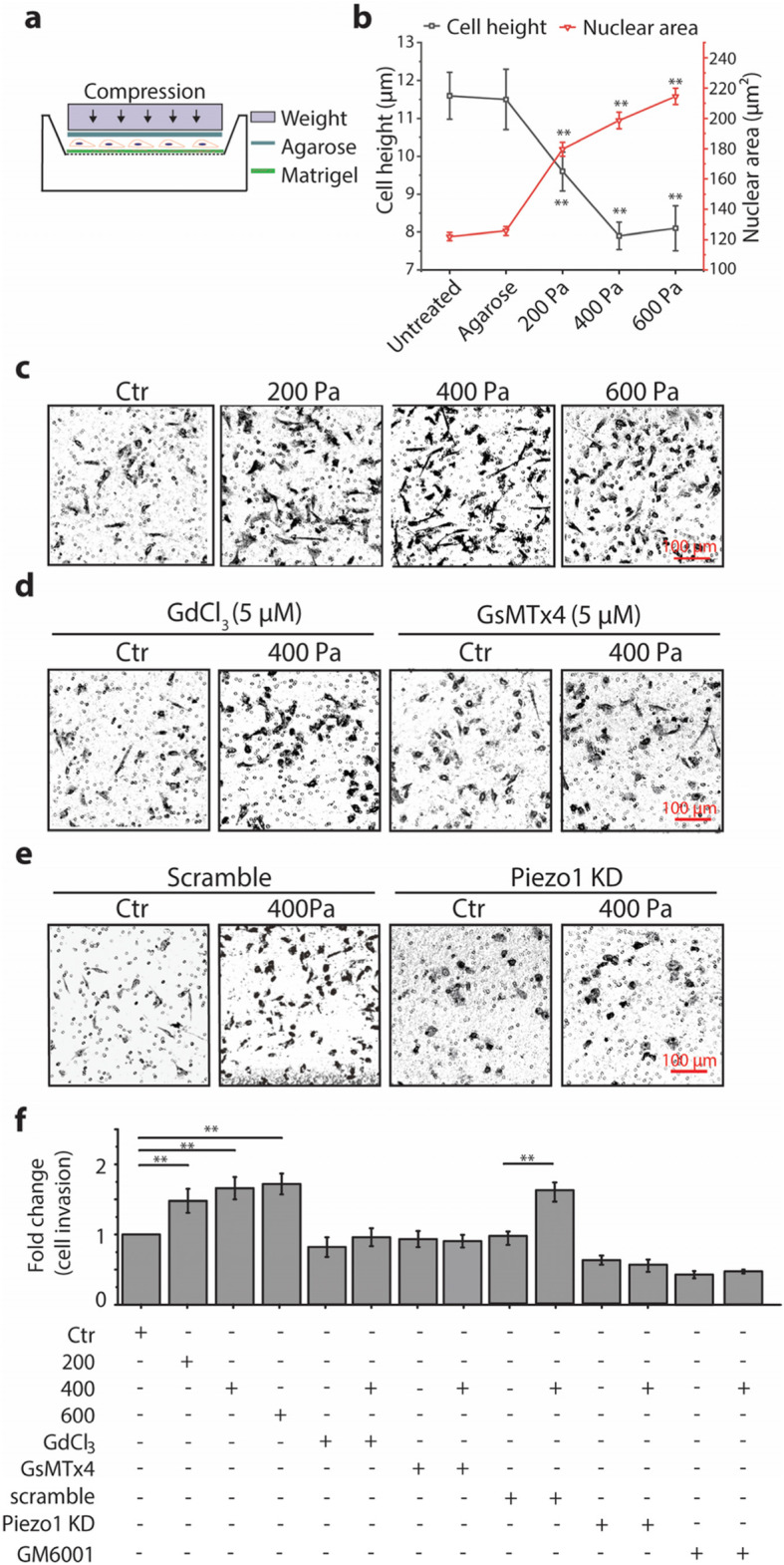
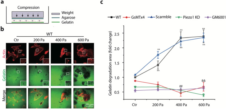
Background: Uncontrolled growth in solid breast cancer generates mechanical compression that may drive the cancer cells into a more invasive phenotype, but little is known about how such compression affects the key events and corresponding regulatory mechanisms associated with invasion of breast cancer cells including cellular behaviors and matrix degradation.

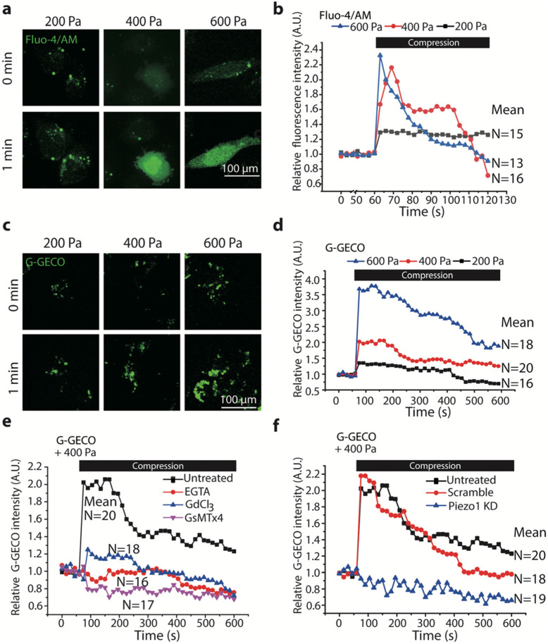
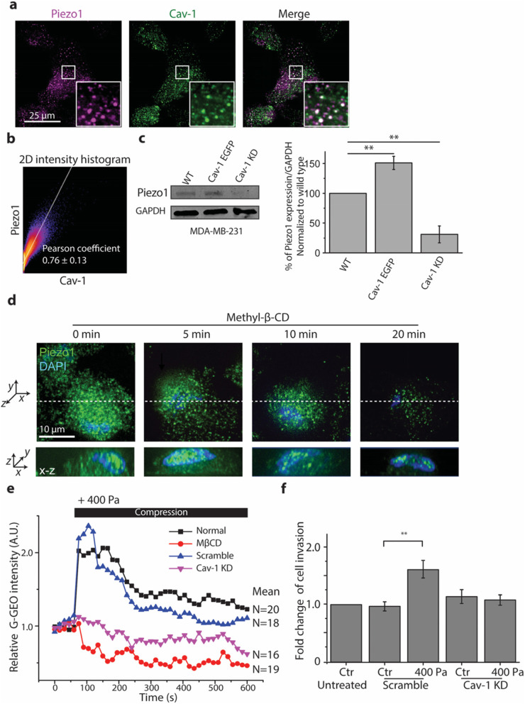
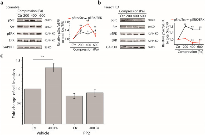
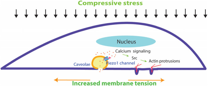
Results: Here we show that compression enhanced invasion and matrix degradation of breast cancer cells. We also identified Piezo1 as the putative mechanosensitive cellular component that transmitted compression to not only enhance the invasive phenotype, but also induce calcium influx and downstream Src signaling. Furthermore, we demonstrated that Piezo1 was mainly localized in caveolae, and both Piezo1 expression and compression-enhanced invasive phenotype of the breast cancer cells were reduced when caveolar integrity was compromised by either knocking down caveolin1 expression or depleting cholesterol content.

Conclusions: Taken together, our data indicate that mechanical compression activates Piezo1 channels to mediate enhanced breast cancer cell invasion, which involves both cellular events and matrix degradation. This may be a critical mechanotransduction pathway during breast cancer metastasis, and thus potentially a novel therapeutic target for the disease.

Keywords: Breast cancer cell; Compression; Invasion; Piezo1.


Conflict of interest statement

The authors declare no competing financial interests in relation to the work described.

Figures

Fig. 1
Fig. 1
Compression enhanced invasion of MDA-MB-231 cells depending on Piezo1. Cell invasion was measured with in vitro transwell invasion assay. a Schematic diagram of the compression experiment using a transwell setup. Cells grown on a membrane filter (8 μm pore) coated with Matrigel for 6 h were covered with 1% of agarose gel and compressed with a specific weight. b The changes of cell height and nuclear area of MDA-MB-231 under compression. Data are presented as means ± s.e.m. n = 4, ** p < 0.01 versus untreated groups. c, d, e Representative images of invaded cells stained with crystal violet under different compression and treated with gadolinium chloride (Gd3+), GsMTx4, or siRNA for Piezo1 under 400 Pa (Bar = 100 μm). f Quantification of the fold change of invaded cells. Data were presented as means ± s.e.m., n = 3, ** p < 0.01 versus control (Ctr) groups
Fig. 2
Fig. 2
Compression promoted matrix degradation in MDA-MB-231 cells. a Schematic diagram of the experiment. Cells grown on a glass-bottom dish coated with FITC-conjugated gelatin for 8 h were covered with 1% of agarose gel and compressed with a specific weight. b Representative images (red: actin, green: gelatin) of compression-promoted gelatin degradation at the ventral side of the cell. Gelatin degradation was visualized by confocal microscopy (60X) as disappearance of green fluorescence. Inset images are magnified views of the boxed regions. c The fold change of gelatin degradation area under different treatment conditions (treated with GsMTx4, Piezo1 KD, or GM6001) as a function of compression normalized to gelatin degradation area at control (Ctr) groups; Data were presented as means ± s.e.m., n = 3, ** p < 0.01 versus control groups, ## and $$ represent p < 0.01 versus 400 Pa and 600 Pa groups in wild type (WT), respectively
Fig. 3
Fig. 3
Compression induced calcium signaling in MDA-MB-231 cells. Representative images of intracellular [Ca2+] (a and c, bar = 100 μm) visualized by confocal microscopy (60X) and time-courses of changing relative mean fluorescence intensity (b and d) of Fluo-4 or G-GECO (normalized to time 0) in MDA-MB-231 cells labeled with Fluo-4/AM or transiently expressing G-GECO before (0 min) and after (1 min) exposure to compression at 200, 400, 600 Pa, respectively. e, f Time-courses of changing relative mean fluorescence intensity of G-GECO in MDA-MB-231 cells pretreated with or without EGTA, Gd3+, GsMTx4, and Piezo1 KD in response to 400 Pa compression. Each experiment assayed 10–20 cells and repeated three times. Black bars in b, d, e, f indicate the period of compression
Fig. 4
Fig. 4
The expression and distribution of Piezo1 in MDA-MB-231 cells were regulated by caveolae. a Representative fluorescence images of Piezo1 (magenta) and caveolae (green) colocalization visualized by confocal microscopy (100X) and 2D intensity histogram output in MDA-MB-231 cells. Insets in both conditions show a magnified view of the boxed regions. b Representative image of 2D intensity histogram output of Coloc2 analysis performed using Fiji software. The text indicates the Pearson coefficient of the pixel-intensity correlation (n = 8). c Western blot images and quantification of Piezo1 expression in wild type (WT), Cav-1 EGFP expressing, and Cav-1 KD MDA-MB-231 cells (means ± s.e.m., n = 3). Cropped images of Western blots are shown and uncropped images are shown in Fig. S8b. ** p < 0.01 versus WT groups. d, Representative fluorescence images of Piezo1 (green) and nucleus (blue) visualized by confocal microscopy (100X) after cells were treated with MβCD for 5 min, 10 min, and 20 min (upper panel: x-y view, lower panel: x-z view, white dashed line shows the position of a section of x-z view). e Time-courses of relative mean fluorescence intensity of G-GECO in MDA-MB-231 cells pretreated with or without MβCD, and Cav-1 KD in response to 400 Pa compression. Each experiment assayed 10–20 cells and repeated three times. The black bar indicates the period of compression. f Quantification of the fold change of invaded cells treated with siRNA for Cav-1 under 400 Pa. Data are presented as means ± s.e.m., n = 3, ** p < 0.01 versus Ctr groups
Fig. 5
Fig. 5
Compression enhanced the activity of Src and ERK. a Western blot analyses of the phosphorylation of Src and ERK in MDA-MB-231 cells pretreated with scramble probes in the absence or presence of compression at 200, 400, 600 Pa. Cropped images of Western blot are shown and uncropped images are shown in Fig. S8d-g. b Western blot analyses of the phosphorylation of Src and ERK in MDA-MB-231 cells pretreated with siRNA for Piezo1 in the absence or presence of compression at 200, 400, 600 Pa. Cropped images of Western blot are shown and uncropped images are shown in Fig. S8h-k. Relative phosphorylation levels were obtained by normalizing to GAPDH expression and value in control (Ctr) groups, n = 3. *p < 0.05 versus Ctr groups; ** p < 0.01 versus Ctr groups. c Quantification of fold change of invaded cells in 400 Pa compression to the Ctr group pretreated with DMSO (vehicle). Data are presented as means ± s.e.m., n = 3, ** p < 0.01 versus Ctr groups
Fig. 6
Fig. 6
Model of compression-promoted invasive phenotype of MDA-MB-231 cells and associated signaling pathways. Together, vertical mechanical compression might increase the lateral plasma membrane tension and activate Piezo1 channels. The opening of Piezo1 mediates the influx of calcium and evokes the downstream signaling pathways such as Src. These activated signaling molecules promote actin protrusions at the ventral side of cells, which in turn mediate enhanced matrix degradation and cell invasion

Similar articles

Cited by

References

    1. Liu AP, Chaudhuri O, Parekh SH. New advances in probing cell-extracellular matrix interactions. Integr Biol (Camb) 2017;9(5):383–405. doi: 10.1039/C6IB00251J. - DOI - PMC - PubMed
    1. Stylianopoulos T, Munn LL, Jain RK. Reengineering the physical microenvironment of tumors to improve drug delivery and efficacy: from mathematical modeling to bench to bedside. Trends Cancer. 2018;4(4):292–319. doi: 10.1016/j.trecan.2018.02.005. - DOI - PMC - PubMed
    1. Petrie RJ, Harlin HM, Korsak LIT, Yamada KM. Activating the nuclear piston mechanism of 3D migration in tumor cells. J Cell Biol. 2016;216(1):93–100. doi: 10.1083/jcb.201605097. - DOI - PMC - PubMed
    1. Wirtz D, Konstantopoulos K, Searson PC. The physics of cancer: the role of physical interactions and mechanical forces in metastasis. Nat Rev Cancer. 2011;11(7):512–522. doi: 10.1038/nrc3080. - DOI - PMC - PubMed
    1. Fernandez-Sanchez ME, Barbier S, Whitehead J, Bealle G, Michel A, Latorre-Ossa H, Rey C, Fouassier L, Claperon A, Brulle L, et al. Mechanical induction of the tumorigenic beta-catenin pathway by tumour growth pressure. Nature. 2015;523(7558):92–95. doi: 10.1038/nature14329. - DOI - PubMed