. 2022 Jan 3;27(1):3.
doi: 10.1186/s11658-021-00299-0.

Mitochondrial potassium channels: A novel calcitriol target

Affiliations

Mitochondrial potassium channels: A novel calcitriol target

Anna M Olszewska et al. Cell Mol Biol Lett. .

Abstract

Background: Calcitriol (an active metabolite of vitamin D) modulates the expression of hundreds of human genes by activation of the vitamin D nuclear receptor (VDR). However, VDR-mediated transcriptional modulation does not fully explain various phenotypic effects of calcitriol. Recently a fast non-genomic response to vitamin D has been described, and it seems that mitochondria are one of the targets of calcitriol. These non-classical calcitriol targets open up a new area of research with potential clinical applications. The goal of our study was to ascertain whether calcitriol can modulate mitochondrial function through regulation of the potassium channels present in the inner mitochondrial membrane.

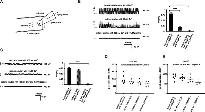
Methods: The effects of calcitriol on the potassium ion current were measured using the patch-clamp method modified for the inner mitochondrial membrane. Molecular docking experiments were conducted in the Autodock4 program. Additionally, changes in gene expression were investigated by qPCR, and transcription factor binding sites were analyzed in the CiiiDER program.

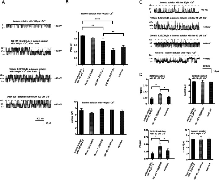
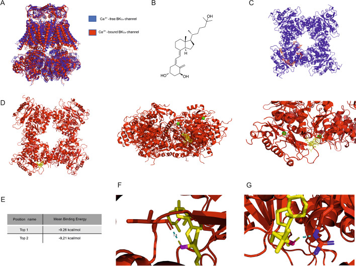
Results: For the first time, our results indicate that calcitriol directly affects the activity of the mitochondrial large-conductance Ca2+-regulated potassium channel (mitoBKCa) from the human astrocytoma (U-87 MG) cell line but not the mitochondrial calcium-independent two-pore domain potassium channel (mitoTASK-3) from human keratinocytes (HaCaT). The open probability of the mitoBKCa channel in high calcium conditions decreased after calcitriol treatment and the opposite effect was observed in low calcium conditions. Moreover, using the AutoDock4 program we predicted the binding poses of calcitriol to the calcium-bound BKCa channel and identified amino acids interacting with the calcitriol molecule. Additionally, we found that calcitriol influences the expression of genes encoding potassium channels. Such a dual, genomic and non-genomic action explains the pleiotropic activity of calcitriol.

Conclusions: Calcitriol can regulate the mitochondrial large-conductance calcium-regulated potassium channel. Our data open a new chapter in the study of non-genomic responses to vitamin D with potential implications for mitochondrial bioenergetics and cytoprotective mechanisms.

Keywords: Calcitriol; Large-conductance calcium-regulated potassium channel; Mitochondria; Patch-clamp.


Conflict of interest statement

The authors declare that they have no competing interests.

Figures

Fig. 1
Fig. 1
Electrophysiological properties of two potassium channels in mitoplasts isolated from glioblastoma and keratinocyte cell line. A Scheme of the patch-clamp experiments performed with calcitriol. B Effect of changed calcium ion concentration and paxilline on the activity of the mitoBKCa channel. (o1) (o2) indicates an open state of channels 1 and 2 respectively. (c) indicates a closed state. C Effect of changed calcium concentration and lidocaine on the activity of the TASK-3 channel. (o) indicates an open state of the channel. (c) indicates a closed state. D, E Resistance of the patch during the external perfusion with calcitriol on mitoplasts from glioblastoma and keratinocyte cells respectively
Fig. 2
Fig. 2
Effect of calcitriol on large-conductance calcium-activated potassium channel from mitochondria isolated from human glioblastoma cells. A Recordings at + 40 mV in symmetrical (150/150 mM) KCl solutions with high calcium ion concentration (100 μM Ca2+); after application of 100 nM calcitriol (1 and 5 min after application), and after wash-out solution with calcitriol. (o1) (o2) indicates an open state of channels 1 and 2 respectively. (c) indicates a closed state. Recordings are from one representative out of three independent experiments. B Bar graphs summarizing the effect of calcitriol on the open probability (Po) of the mitoBKCa channel and the current amplitude (pA) in the presence of 100 μM calcium. The upper panel shows changes of the Po after adding increasing concentrations of calcitriol and wash-out (n = 3). The lower panel shows changes of the current after adding increasing concentrations of calcitriol and wash-out (n = 3). C Recordings at + 40 mV in symmetrical (150/150 mM) KCl solutions with low calcium ion concentration (10 μM Ca2+, control), after application of 100 nM and 300 nM calcitriol and wash-out. (o1) (o2) indicates an open state of channels 1 and 2 respectively. (c) indicates a closed state. Recordings are from one representative out of three independent experiments. D Bar graphs summarizing the effect of 100 nM and 300 nM calcitriol and wash-out on the open probability (Po) of the mitoBKCa channel and the current amplitude (pA) in 10 μM Ca2+ ion concentration. The left panel shows changes of the Po after adding 100 nM or 300 nM calcitriol and wash-out (n = 3). The right panel shows changes in the current after adding 100 nM or 300 nM calcitriol and wash-out (n = 3)
Fig. 3
Fig. 3
Effect of calcitriol on two-pore domain potassium channels from mitochondria isolated from human keratinocytes. A Recordings at + 40 mV in symmetrical (150/150 mM) KCl solutions with high calcium ion concentration (100 μM Ca2+, control) and low calcium concentration (10 μM Ca2+, control) and after application of 100 nM calcitriol. (o) indicates an open state of the channel. (c) indicates a closed state. Recordings are from one representative out of three independent experiments. B Bar graphs summarizing the effect of calcitriol on the open probability (Po) of the mitoTASK-3 channel and current amplitude (pA) in the presence of high and low calcium conditions. The left panel shows changes of Po after adding 30 nM, 100 nM, 300 nM calcitriol in the presence of high and low Ca2+ ion concentration (n = 3). The right panel shows changes in the current after adding 30 nM, 100 nM, 300 nM calcitriol in the presence of high and low Ca2+ ion concentration (n = 3)
Fig. 4
Fig. 4
Calcitriol effect on expression level of potassium channels. A Calcitriol effect on expression of calcium-regulated potassium channels (KCNN1-KCNN4; KCNMA1) and two pore-domain potassium channel TASK-3 (KCNK9) in HaCaT cells. B JASPAR matrices are used in CiiiDER program analysis. C Prediction of VDR binding sites in the BK channel gene (KCNMA1) by the CiiiDER program. VDR binding sites (JASPAR matrix MA0693.2) in KCNMA1 sequence 10 kb upstream of ATG were marked in black. VDR binding site (JASPAR matrix MA0074.1) in KCNMA1 sequence 10 kb upstream of ATG was marked in grey
Fig. 5
Fig. 5
Predicted docking poses of calcitriol on the BKCa channel with and without Ca2+and a close-up of the specific residues predicted to interact with calcitriol. A Protein alignments of two structures in 3D, BKCa channel Ca2+-bound (red color; PDB: 6v22) and BKCa channel Ca2+-free (blue color; PDB: 6v35). B calcitriol molecule C Docking poses of calcitriol (red color) on the cytoplasmic domain of the Ca2+-free BKCa channel, showing wide diversity of binding poses. D Predicted docking poses of calcitriol (yellow color) on the cytoplasmic domain of the Ca2+-bound (green spheres) BKCa channel. E The predicted binding energies of two calcitriol binding poses (top 1, top 2). F Close-up of the specific residues of the BKCa channel protein predicted to interact with calcitriol (top 1) hydroxyl group on C1 calcitriol carbon. G Close-up of the specific residues of the BKCa channel protein predicted to interact with calcitriol (top 1) hydroxyl group on C25 calcitriol carbon

Similar articles

Cited by

References

    1. Slominski AT, Chaiprasongsuk A, Janjetovic Z, Kim TK, Stefan J, Slominski RM, et al. Photoprotective properties of vitamin D and lumisterol hydroxyderivatives. Cell Biochem Biophys. 2020;78(2):165–180. - PMC - PubMed
    1. Slominski AT, Brożyna AA, Zmijewski MA, Jóźwicki W, Jetten AM, Mason RS, et al. Vitamin D signaling and melanoma: role of vitamin D and its receptors in melanoma progression and management. Lab Invest. 2017;97(6):706–724. - PMC - PubMed
    1. Slominski AT, Brożyna AA, Zmijewski MA, Janjetovic Z, Kim TK, Slominski RM, et al. The role of classical and novel forms of vitamin D in the pathogenesis and progression of nonmelanoma skin cancers. Adv Exp Med Biol. 2020;1268:257–283. - PMC - PubMed
    1. Chen J, Tang Z, Slominski AT, Li W, Żmijewski MA, Liu Y. Vitamin D and its analogs as anticancer and anti-inflammatory agents. Eur J Med Chem. 2020;207:112738. - PubMed
    1. Zmijewski MA, Carlberg C. Vitamin D receptor(s): in the nucleus but also at membranes? Exp Dermatol. 2020;29(9):876–884. - PubMed

MeSH terms

Substances