. 2022 Jan 3;22(1):1.
doi: 10.1186/s12935-021-02402-z.

SCUBE3 downregulation modulates hepatocellular carcinoma by inhibiting CCNE1 via TGFβ/PI3K/AKT/GSK3β pathway

Affiliations

SCUBE3 downregulation modulates hepatocellular carcinoma by inhibiting CCNE1 via TGFβ/PI3K/AKT/GSK3β pathway

Pan Xu et al. Cancer Cell Int. .

Abstract

Objectives: We aimed to verify the role of signal peptide-CUB-EGF-like domain-containing protein3 (SCUBE3) in the hepatocellular carcinoma (HCC) progression.

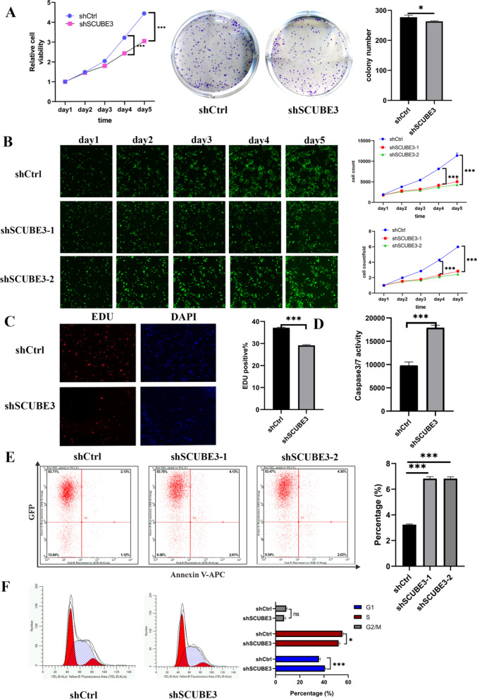
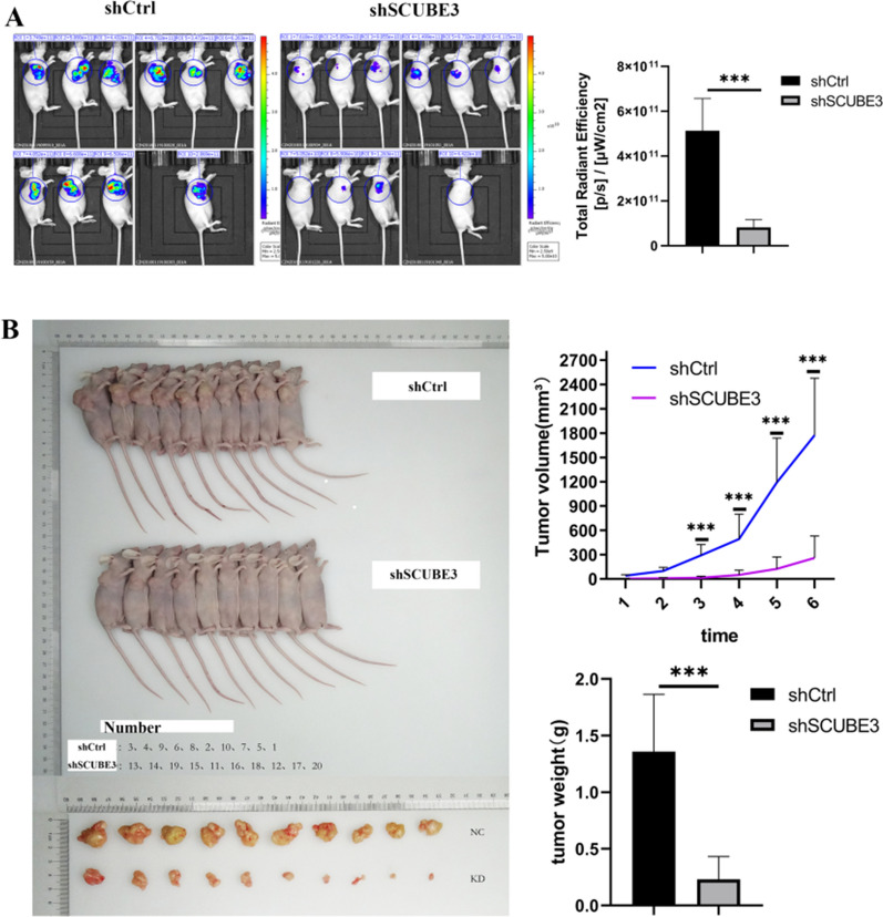
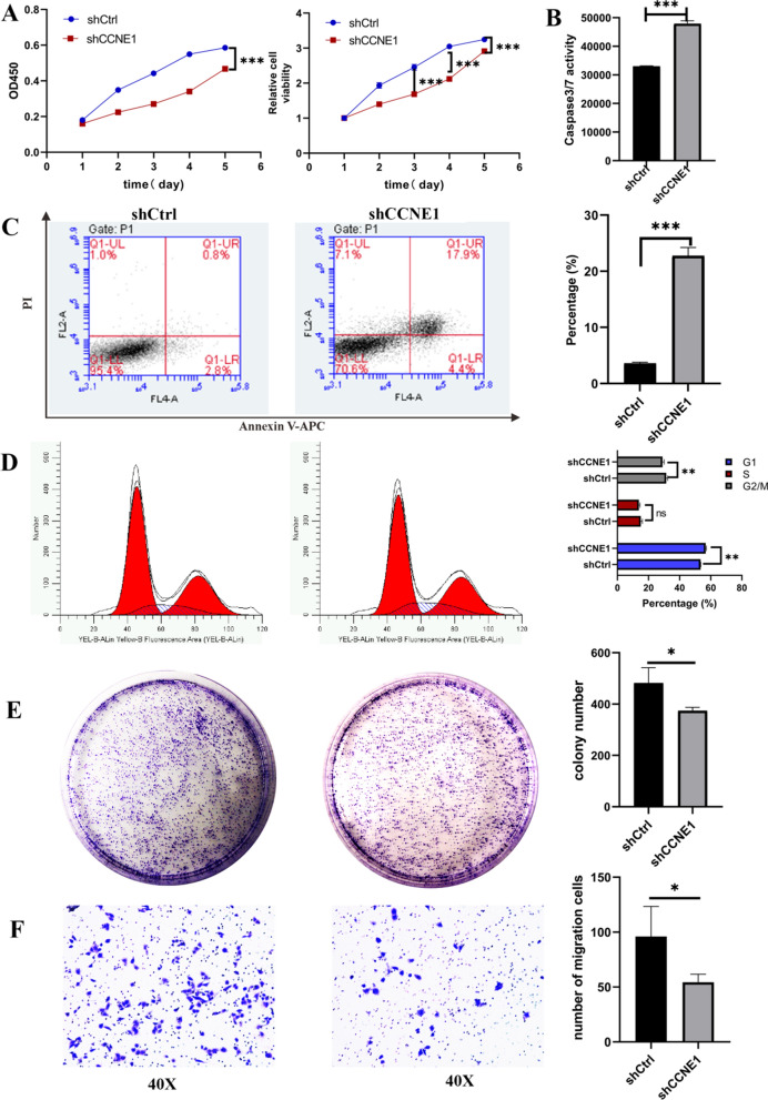
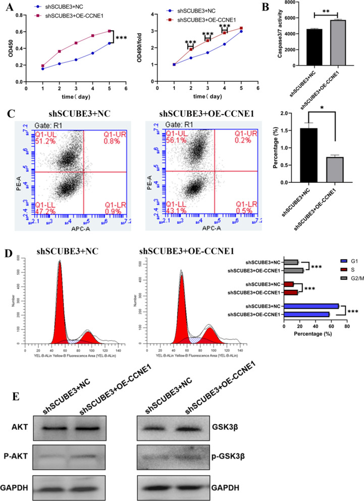
Methods: The role of SCUBE3 in HCC cell proliferation, apoptosis, and cell cycle in vitro were detected using MTT assay, colony formation assay, 5-ethynyl-2´-deoxyuridine assay (EDU), Celigo cell counting assay, Caspase3/7 activity assay, and flow cytometry. The effect of SCUBE3 on HCC cell proliferation in vivo was inspected by a xenograft tumour model in nude mice. The related mechanisms were further studied.

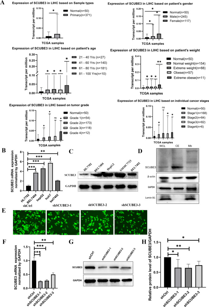
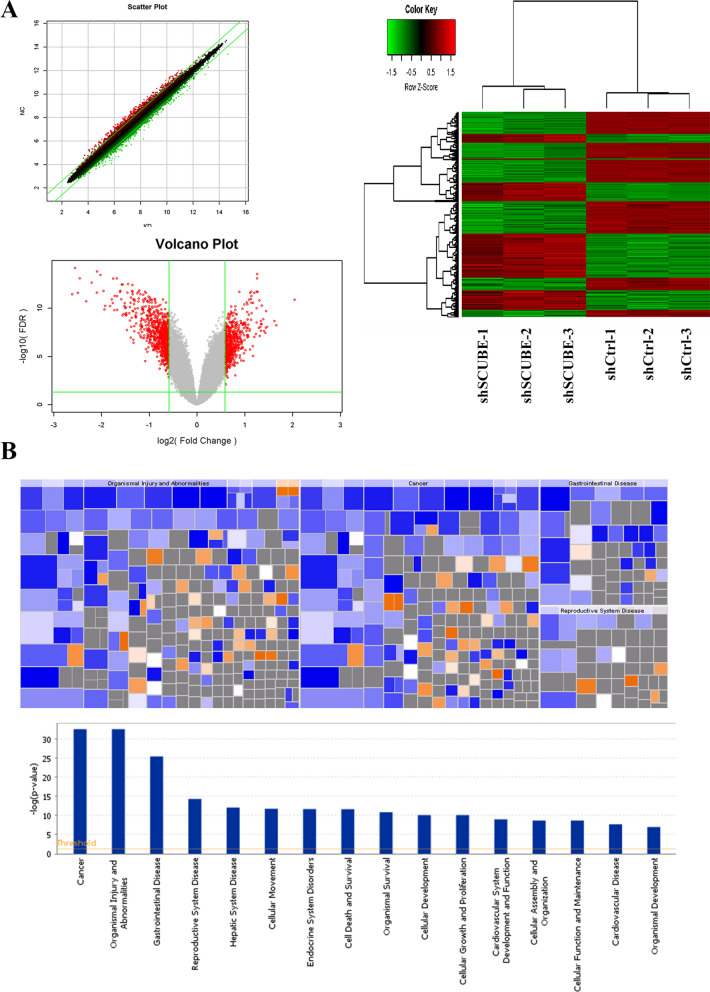
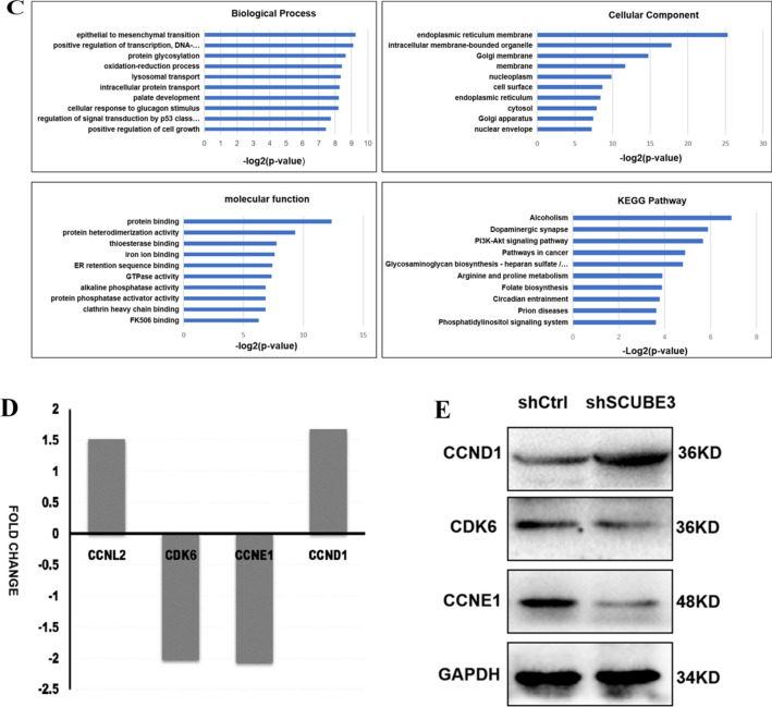
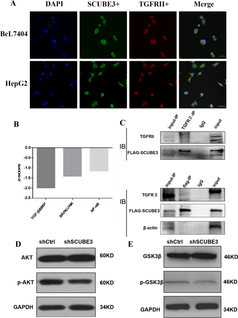
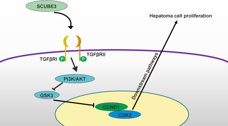
Results: The level of SCUBE3 was upregulated in HCC tissues and cell lines. Knockdown of SCUBE3 inhibited proliferation, promoted apoptosis, and induced cell cycle arrest in HCC cell lines in vitro and in vivo. Screening of cell cycle-related proteins revealed that CCNL2, CDK6, CCNE1, and CCND1 exhibited a significantly different expression profile. We found that SCUBE3 may promote the proliferation of HCC cells by regulating CCNE1 expression. The pathway enrichment analysis showed that the TGFβ signalling pathway and the PI3K/AKT signalling pathway were significantly altered. Co-immunoprecipitation results showed that SCUBE3 binds to the TGFβRII receptor. SCUBE3 knockdown inhibited the PI3K/AKT signalling pathway and the phosphorylation of GSK3β to inhibit its kinase activity.

Conclusions: SCUBE3 promotes HCC development by regulating CCNE1 via TGFβ/PI3K/AKT/GSK3β pathway. In addition, SCUBE3 may be a new molecular target for the clinical diagnosis and treatment of HCC.

Keywords: AKT; CCNE1; Cell proliferation; Hepatocellular carcinoma; SCUBE3.


Conflict of interest statement

The authors declare that they have no competing interests.

Figures

Fig. 1
Fig. 1
Expression analysis of SCUBE3 and establishment and validation of scube3 knockdown cell model. A SCUBE3 expression in tumour and normal tissues in the TCGA database from UALCAN database. SCUBE3 expression is based on the pa'tients sex, age, body weight, tumour grade and cancer stage. B, C RT-qPCR and western blot analysis of SCUBE3 mRNA and protein expression in normal hepatocytes HL7702 and four HCC cell lines (Bel7404, Bel7402, HepG2, and SMMC7721). D Western blotting was used to detect the expression level of SCUBE3 in the whole protein, cytoplasmic protein, and membrane protein samples of Bel7404 HCC cells. E Bel7404 cells were transfected with control shRNA (shCtrl) and three shSCUBE3 lentiviruses (shSCUBE3-1, shSCUBE3-2, shSCUBE3-3). Green fluorescence protein (GFP) expression illustrated to transfect with lentivirus successfully. F The expression levels of SCUBE3 in the cells were determined through reverse-transcriptase PCR. GH The protein expression of SCUBE3 was examined by western blotting (n = 3, ns,no significance,*p < 0.05,**p < 0.01, and ***p < 0.001 compared with normal cells)
Fig. 2
Fig. 2
Knockdown of SCUBE3 expression suppressed proliferation, promoted apoptosis, and induced cell cycle arrest in HCC cells. A MTT-assays were performed to evaluate the viability of negative control and cells expressing low levels of SCUBE3, and Colony formation assay using knockdown of SCUBE3 cells. B The proliferation rate of Bel7404 cells was reduced by SCUBE3 knockdown, which was observed using the Celigo cell counting assay. C EdU assays were used to determine the effect of SCUBE3 on the proliferation of Bel7404 cells. D Relative caspase activity measured by caspase 3/7 assay showed apoptosis in knockdown of SCUBE3 cells (shSCUBE3). E Effect of SCUBE3 on apoptosis was analyzed through flow cytometry using Annexin V-APC staining. F Cell cycle distribution after inhibition of SCUBE3 was analyzed through flow cytometry (n = 3,*p < 0.05, **p < 0.01, ***p < 0.001. ns, no significance, KD, knockdown, NC, negative control)
Fig. 3
Fig. 3
SCUBE3 promotes HCC cell proliferation in vivo. A In vivo imaging of subcutaneous tumour formation in nude mice implanted with SCUBE3 knockdown and control cells. Quantification of fluorescence values of the two groups. B Photographs of tumours taken after four weeks. Comparison of tumour volume and weight of SCUBE3 knockdown cells and control cells after subcutaneous tumour formation in nude mice (n = 10, The results represent the mean ± SD,***p < 0.001)
Fig. 4
Fig. 4
Affymetrix GeneChip microarray analysis and validation. A Analysis of significant differences according to the screening criteria of fold change |> 1.5| and false discovery rate (FDR) < 0.05. Scatter plot, volcano plot, heat map were constructed based on the genes differentially expressed between the control and SCUBE3-knockdown cells. B Disease and functional enrichment analysis of differential genes by IPA. The heat map of disease and function showed the relationship between the activation and inhibition of disease and function by the change of differential gene expression level. Orange indicates that the disease or functional state is activated (Z-score > 0), blue indicates that the disease or functional state is inhibited (Z-score < 0), and grey indicates that the disease or functional state is not determined (Z-score cannot be calculated). According to the internal algorithm and standard of IPA, Z-score > 2 means that the disease or function is significantly activated, and Z-score <—2 means that the disease or function is significantly inhibited. The diseases or functions significantly activated include Organic death (Z-score = 3.247), morbidity or mortality (Z-score = 3.200), etc.The diseases or functions significantly inhibited included: viral infection (Z-score = − 3.110), invasion of cells (Z-score = − 2.933). Histogram of disease and functional enrichment analysis statistics. C Significant GO terms associated with SCUBE3 knockdown in HCC cells.Analysis of cellular component (GO-CC), Analysis of biological process (GO-BP). Analysis of molecular function (GO-MF). KEGG Pathway enrichment. D Four genes associated with cell cycle regulation were screened by the microarray Chip analysis following the screening criteria. E The expression levels of three proteins (CCNE1, CCND1, CDK6) were verified by WB
Fig. 4
Fig. 4
Affymetrix GeneChip microarray analysis and validation. A Analysis of significant differences according to the screening criteria of fold change |> 1.5| and false discovery rate (FDR) < 0.05. Scatter plot, volcano plot, heat map were constructed based on the genes differentially expressed between the control and SCUBE3-knockdown cells. B Disease and functional enrichment analysis of differential genes by IPA. The heat map of disease and function showed the relationship between the activation and inhibition of disease and function by the change of differential gene expression level. Orange indicates that the disease or functional state is activated (Z-score > 0), blue indicates that the disease or functional state is inhibited (Z-score < 0), and grey indicates that the disease or functional state is not determined (Z-score cannot be calculated). According to the internal algorithm and standard of IPA, Z-score > 2 means that the disease or function is significantly activated, and Z-score <—2 means that the disease or function is significantly inhibited. The diseases or functions significantly activated include Organic death (Z-score = 3.247), morbidity or mortality (Z-score = 3.200), etc.The diseases or functions significantly inhibited included: viral infection (Z-score = − 3.110), invasion of cells (Z-score = − 2.933). Histogram of disease and functional enrichment analysis statistics. C Significant GO terms associated with SCUBE3 knockdown in HCC cells.Analysis of cellular component (GO-CC), Analysis of biological process (GO-BP). Analysis of molecular function (GO-MF). KEGG Pathway enrichment. D Four genes associated with cell cycle regulation were screened by the microarray Chip analysis following the screening criteria. E The expression levels of three proteins (CCNE1, CCND1, CDK6) were verified by WB
Fig. 5
Fig. 5
CCNE1 silence inhibit Bel7404 cell proliferation, migration, arrested G1/S transition and promote apoptosis. A 5 days of continuous MTT assays. B Knockdown of CCNE1 remarkably enhanced caspase-3/7 activity in the shCCNE1 group. C Knockdown of CCNE1 promoted cell apoptosis. D Analysis of cell cycle profiles after CCNE knockdown in Bel7404 cells. E Colony formation assay using cells infected with CCNE1 lentiviral vector and empty vectors. Representative images were acquired, and the colonies were counted after two weeks. F The inhibition effect of CCNE1 silence on cell migration was assessed by transwell assay. After 24 h of incubation, the cells were fixed and stained. Representative images of transwell chamber assays were acquired, and quantification of the number of cells which migrated through the basement membrane (n = 5 per group) (*p < 0.01, **p < 0.01,***p < 0.001 compared with shCtrl)
Fig. 6
Fig. 6
Overexpression of CCNE1 in SCUBE3 knockdown Bel7404 cells regulates proliferation and apoptosis. A MTT proliferation assay. B caspase 3/7 activity assay. C Detection of apoptosis in cells with both SCUBE3 knockdown and CCNE1 overexpression and control cells by the Annexin V-APC/PI double staining assay and, D cell cycle analysis of cells with both SCUBE3 knockdown and CCNE1 overexpression and control cells. E SCUBE3 knockdown cells were treated with overexpression lentivirus of CCNE1 for 72 h, Cell lysates were analyzed by immunoblotting using the indicated antibodies (*p < 0.01, **p < 0.01,***p < 0.001)
Fig. 7
Fig. 7
SCUBE3 binds to the TGFβIIR. A Co-localization of SCUBE3 and TGFβIIR. HepG2 and Bel7404 cells were stained with antibodies against SCUBE3 and TGFβIIR, followed by incubation with FITC-conjugated donkey anti-rat or anti-mouse IgG. The cells were visualized using a confocal microscope. The yellow areas represent protein co-localization. B The TGFβ pathway is significantly downregulated after SCUBE3 knockdown. C Co-immunoprecipitation assay results indicate that SCUBE3 interacts with TGFβIIR. D Western blot analysis of AKT and p-AKT expression in SCUBE3 knockdown cells and control cells. E Western blot analysis of GSK3β and p-GSK3β expression in SCUBE3 knockdown cells and control cells
Fig. 8
Fig. 8
Schematic diagram of the validation hypothesis of this study: SCUBE3 activates the PI3K/AKT pathway by binding to the TGFβRII receptor to phosphorylate GSK3β to inhibit its kinase activity, thereby inhibiting the ubiquitination of CCNE1, leading to the accumulation of CCNE1 and ultimately promoting the proliferation of liver cancer cells

References

    1. Villanueva A. Hepatocellular carcinoma. N Engl J Med. 2019;380(15):1450–1462. - PubMed
    1. Ozturk M, Batur T, Ekin U, et al. Molecular pathogenesis of liver cancer. J Gastrointest Cancer. 2017;48(3):222–224. - PubMed
    1. Zender L, Villanueva A, Tovar V, Sia D, Chiang DY, Llovet JM. Cancer gene discovery in hepatocellular carcinoma. J Hepatol. 2010;52(6):921–929. - PMC - PubMed
    1. Thompson MD, Monga SP. WNT/beta-catenin signaling in liver health and disease. Hepatology. 2007;45(5):1298–1305. - PubMed
    1. Min L, He B, Hui L. Mitogen-activated protein kinases in hepatocellular carcinoma development. Semin Cancer Biol. 2011;21(1):10–20. - PubMed