. 2022 Jan 3;41(1):3.
doi: 10.1186/s13046-021-02208-x.

Epigenetic regulation of ferroptosis via ETS1/miR-23a-3p/ACSL4 axis mediates sorafenib resistance in human hepatocellular carcinoma

Affiliations

Epigenetic regulation of ferroptosis via ETS1/miR-23a-3p/ACSL4 axis mediates sorafenib resistance in human hepatocellular carcinoma

Yuanjun Lu et al. J Exp Clin Cancer Res. .

Abstract

Background: Drug resistance to sorafenib greatly limited the benefits of treatment in patients with hepatocellular carcinoma (HCC). MicroRNAs (miRNAs) participate in the development of drug resistance. The key miRNA regulators related to the clinical outcome of sorafenib treatment and their molecular mechanisms remain to be identified.

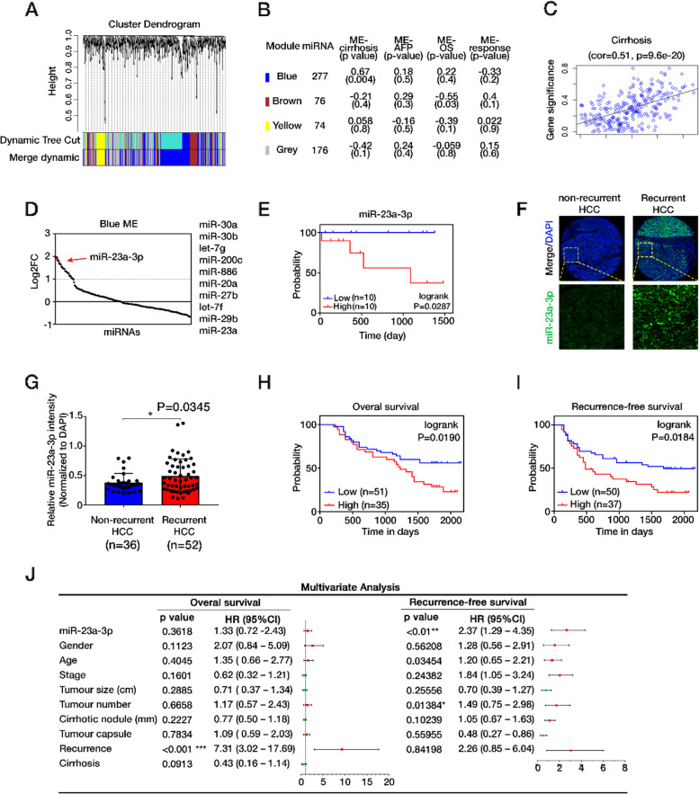
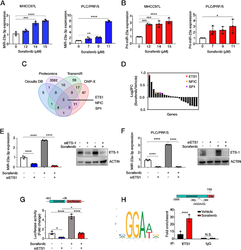
Methods: The clinical significance of miRNA-related epigenetic changes in sorafenib-resistant HCC was evaluated by analyzing publicly available databases and in-house human HCC tissues. The biological functions of miR-23a-3p were investigated both in vitro and in vivo. Proteomics and bioinformatics analyses were conducted to identify the mechanisms that regulating miR-23a-3p. Luciferase reporter assay and chromatin immunoprecipitation (ChIP) assay were used to validate the binding relationship of miR-23a-3p and its targets.

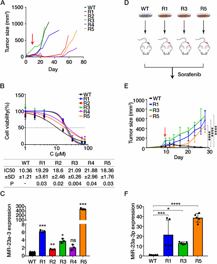
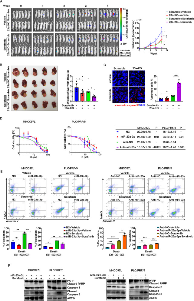
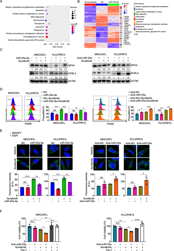
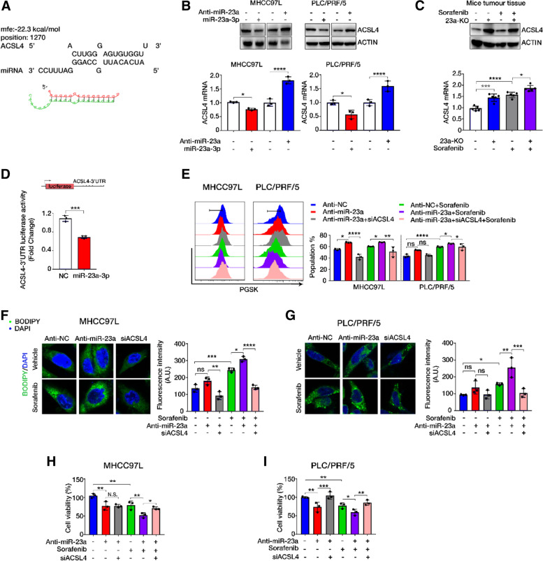
Results: We found that miR-23a-3p was the most prominent miRNA in HCC, which was overexpressed in sorafenib non-responders and indicated poor survival and HCC relapse. Sorafenib-resistant cells exhibited increased miR-23a-3p transcription in an ETS Proto-Oncogene 1 (ETS1)-dependent manner. CRISPR-Cas9 knockout of miR-23a-3p improved sorafenib response in HCC cells as well as orthotopic HCC tumours. Proteomics analysis suggested that sorafenib-induced ferroptosis was the key pathway suppressed by miR-23a-3p with reduced cellular iron accumulation and lipid peroxidation. MiR-23a-3p directly targeted the 3'-untranslated regions (UTR) of ACSL4, the key positive regulator of ferroptosis. The miR-23a-3p inhibitor rescued ACSL4 expression and induced ferrotoptic cell death in sorafenib-treated HCC cells. The co-delivery of ACSL4 siRNA and miR-23a-3p inhibitor abolished sorafenib response.

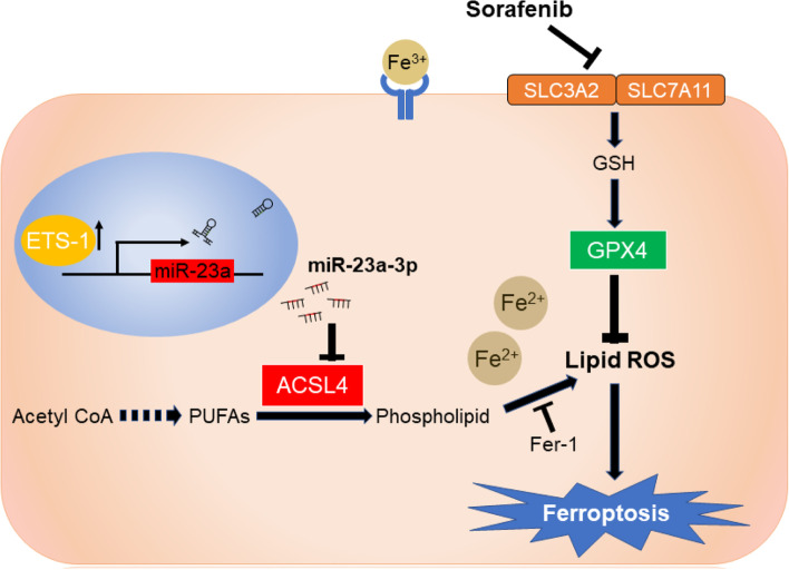
Conclusion: Our study demonstrates that ETS1/miR-23a-3p/ACSL4 axis contributes to sorafenib resistance in HCC through regulating ferroptosis. Our findings suggest that miR-23a-3p could be a potential target to improve sorafenib responsiveness in HCC patients.

Keywords: ACSL4; ETS1; Ferroptosis; Hepatocellular carcinoma; MiR-23a-3p; Sorafenib resistance.


Conflict of interest statement

The authors declare that they have no competing interests

Figures

Fig. 1
Fig. 1
The evaluation of clinical importance of miR-23a-3p. A Dendrogram of miRNAs clustered based on a dissimilarity measure (1-TOM) through WGCNA analysis. B Module member count, ME-clinical traits correlation (Pearson) and P-value indicated for each module. C Scatterplots showing the correlations between gene module membership in the blue module and gene significance for cirrhosis. D A dot plot showing the ranking of miRNAs according to their log2 fold change. E Progression-free survival curve of miR-23a-3p in HCC patients treated with sorafenib. Data were gathered from the GSE56059. F Representative images of miR-23a-3p and DAPI staining in the human HCC tissue microarray. G Increased miR-23a-3p expression observed in recurrent HCC, P = 0.0345. H OS and I RFS outcomes based on Kaplan-Meier survival analysis. The increased expression of miR-23a-3p was significantly associated with poor OS and DFS with P-values 0.0190 and 0.0184, respectively. F Multivariate Cox analysis showing the association between clinicopathologic factors and HCC patient survival outcome. The expression of MiR-23a-3p is significant only in RFS. **P < 0.01, *P < 0.05
Fig. 2
Fig. 2
Upregulated miR-23a-3p in in vivo-generated sorafenib resistant HCC cells. A Tumour growth of mice was recorded every 3 days. WT: vehicle group; R1–5: sorafenib-treated group. B Data are represented as the percentage of WT and R1-R5 cells, and each of the experiment was performed in triplicate. The IC50 values of sorafenib in tumour cells for 24 h were determined by MTT assay and were calculated by GraphPad Prism 7 with the equation of Y=Bottom+(Top-Bottom)/(1 + 10^((LogIC50X)*HillSlope)). Mean ± SD of IC50 was displayed and P-value of comparison between WT and sorafenib resistance group was shown. C The expression of miR-23a-3p in both parental and in vivo-generated sorafenib resistant cell lines. D and E Tumour growth in the re-injected mouse models. WT: mice with parental cells; R1/3/5: mice with in vivo-generated sorafenib resistant cells. F The expression of miR-23a-3p in the re-injected mouse model. The arrow indicates the start of sorafenib administration. One-way ANOVA, *P < 0.05, **P < 0.01, ***P < 0.005, ****P < 0.0001
Fig. 3
Fig. 3
ETS1 directly stimulates miR-23a-3p transcription upon sorafenib treatment. A The expression of miR-23a-3p and B pri-miR-23a in MHCC97L and PLC/PRF/5 cells treated with sorafenib for 24 h. The doses of sorafenib were corresponding to 0, IC10, IC15, and IC20 values of sorafenib. C Venn diagram showing the intersections of data from proteomics analysis, ChIP-X, TransmiR and Circuit. Three TFs: ETS1, NFIC, and SP1 were the common TFs. D Gene ranking of potential TFs of proteomics data according to their log2FC. ETS1 was the highest upregulated TF. E The expression of miR-23a-3p in MHCC97L and F PLC/PRF/5 by qRT-PCR and the expression of ETS1 by immunoblotting. G Luciferase activity of pGL-23AP639 in HEK293. One-way ANOVA, p < 0.005***, p < 0.0001****. H The binding motif of ETS-1 on miR-23a promoter and the fold enrichment of fragments of miR-23a promoter was higher in sorafenib treated MHCC97L after the pulldown by ETS-1 Ab. The IgG Ab group was set as the negative control. Unpair t-test, p < 0.0001****
Fig. 4
Fig. 4
Suppression of miR-23a-3p potentiated sorafenib response both in vivo and in vitro. A Mice with orthotopic implantation of Scramble and 23a-KO cells (n = 6) and the signal intensity in HCC in 4 weeks. B Images and the weight of HCC-bearing livers. Yellow circles indicate HCC tumours. C Cleaved caspase-3 staining in the orthotopic HCC section. Cells with green staining are apoptotic cells. D The IC50 value of sorafenib after miR-23a-3p and Anti-miR-23a transfection in MHCC97L and PLC for 24 h. Mean ± SD of IC50 was displayed and P-value of comparison between groups was shown. E and F The effect of miR-23a-3p in cell apoptosis determined by FACS analysis using Annexin V-FITC/7-AAD double staining kit and immunoblotting
Fig. 5
Fig. 5
MiR-23a-3p suppressed sorafenib-induced ferroptosis. A Dot plot showing KEGG pathway enrichment for differentially expressed proteins among NC + sorafenib, NC, and miR-23a-3p groups. B Heatmap showing protein expression pattern with KEGG pathway annotation. C The protein expression of GPX4 and ACSL4. D Chelatable iron accumulation was detected by fluorescent indicator Phen Green SK with dynamic quenching signals. E The deposition of lipid peroxides was stained with BODIPY and measured by LSM780 confocal imaging. F Cell viability of HCC cells examined by MTT assay
Fig. 6
Fig. 6
ACSL4 was targeted by miR-23a-3p. A Predicted miR-23a-3p binding sites in the 3’UTR of ACSL4 mRNA according to the computational algorithms of RNA hybrid. B The mRNA and protein expression of ACSL4 in HCC cell lines after transfection with miR-23a-3p mimics and Anti-miR-23a for 24 h. C ACSL4 expression in the orthotopic HCC tissues showing high ACSL4 in the 23a-KO group. D Luciferase activity of ACSL4 3’UTR after miR-23a-3p mimics transfection in HEK239 cells. E ACSL4 siRNA neutralized the induced accumulation of chelatable iron by Anti-miR-23a upon sorafenib treatment. F and G ACSL4 siRNA inhibited the deposition of lipid peroxides increased by miR-23a-3p inhibitor upon sorafenib treatment. H and I Suppression of cell viability by miR-23a-3p inhibitor under sorafenib treatment reversed by ACSL4 siRNA co-transfection
Fig. 7
Fig. 7
Schematic model of the mechanism underlying miR-23a-3p on sorafenib resistance in HCC. Sorafenib treatment triggered ferroptosis via lipid ROS production and chelatable iron accumulation. The ETS1 upregulated by sorafenib was a key transcription factor of miR-23a-3p that directly enhanced miR-23a-3p expression. MiR-23a-3p recognized and bound to ACSL4 3’UTR to limit lipid ROS production, thus attenuating sorafenib-induced ferroptotic cell death in HCC

References

    1. Siegel RL, Miller KD, Fuchs HE, Jemal A. Cancer statistics, 2021. CA Cancer J Clin. 2021;71(1):7–33. - PubMed
    1. Villanueva A. Hepatocellular Carcinoma. N Engl J Med. 2019;380(15):1450–1462. - PubMed
    1. Llovet JM, Ricci S, Mazzaferro V, Hilgard P, Gane E, Blanc JF, de Oliveira AC, Santoro A, Raoul JL, Forner A, et al. Sorafenib in advanced hepatocellular carcinoma. N Engl J Med. 2008;359(4):378–390. - PubMed
    1. Tang W, Chen Z, Zhang W, Cheng Y, Zhang B, Wu F, Wang Q, Wang S, Rong D, Reiter FP, et al. The mechanisms of sorafenib resistance in hepatocellular carcinoma: theoretical basis and therapeutic aspects. Signal Transduct Target Ther. 2020;5(1):87. - PMC - PubMed
    1. Tang W, Chen Z, Zhang W, Cheng Y, Zhang B, Wu F, Wang Q, Wang S, Rong D, Reiter F. The mechanisms of sorafenib resistance in hepatocellular carcinoma: theoretical basis and therapeutic aspects. Signal Transduct Target Ther. 2020;5(1):1–15. - PMC - PubMed