. 2022 May;126(9):1329-1338.
doi: 10.1038/s41416-021-01667-5. Epub 2022 Jan 3.

Prognostic implications of adaptive immune features in MMR-proficient colorectal liver metastases classified by histopathological growth patterns

Affiliations

Prognostic implications of adaptive immune features in MMR-proficient colorectal liver metastases classified by histopathological growth patterns

Nouredin Messaoudi et al. Br J Cancer. 2022 May.

Abstract

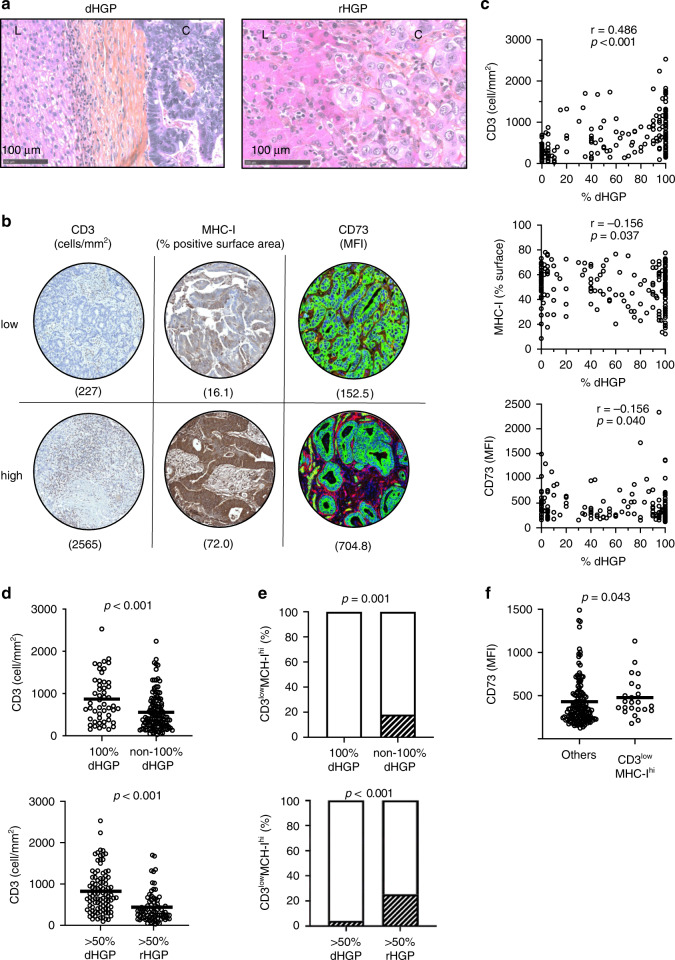
Background: After resection, colorectal cancer liver metastases (CRLM) surrounded by a desmoplastic rim carry a better prognosis than the metastases replacing the adjacent liver. However, these histopathological growth patterns (HGPs) are insufficient to guide clinical decision-making. We explored whether the adaptive immune features of HGPs could refine prognostication.

Methods: From 276 metastases resected in 176 patients classified by HGPs, tissue microarrays were used to assess intratumoral T cells (CD3), antigen presentation capacity (MHC class I) and CD73 expression producing immunosuppressive adenosine. We tested correlations between these variables and patient outcomes.

Results: The 101 (57.4%) patients with dominant desmoplastic HGP had a median recurrence-free survival (RFS) of 17.1 months compared to 13.3 months in the 75 patients (42.6%) with dominant replacement HGP (p = 0.037). In desmoplastic CRLM, high vs. low CD73 was the only prognostically informative immune parameter and was associated with a median RFS of 12.3 months compared to 26.3, respectively (p = 0.010). Only in dominant replacement CRLM, we found a subgroup (n = 23) with high intratumoral MHC-I expression but poor CD3+ T cell infiltration, a phenotype associated with a short median RFS of 7.9 months.

Conclusions: Combining the assessments of HGP and adaptive immune features in resected CRLM could help identify patients at risk of early recurrence.


Conflict of interest statement

JS is a permanent member of the scientific advisory board of Surface Oncology and holds stocks of Surface Oncology. ST is receiving non-clinical research funding from Bristol-Myers Squibb, clinical research funding from GlaxoSmithKline, Iovance Biotherapeutics and Turnstone Biologics. All other authors report no conflicts of interest concerning this specific publication.

Figures

Fig. 1
Fig. 1. Histopathological growth patterns and immune features assessed in colorectal liver metastasis.
a Representative hematoxylin and eosin (H&E)-stained tissue sections of the interface between the cancer cells (C) of the metastasis and the adjacent non-tumoral liver (L). A fibrous band is found at the interface of the metastasis with desmoplastic histopathological growth pattern (dHGP, left), whereas cancer cells grow in the liver cell plates by replacing the hepatocytes in the replacement type (rHGP, right). b Representative tissue microarray cores of metastases with low and high CD3+ T cell infiltration (DAB-brown signal), MHC-I percent surface expression (DAB-brown signal) and mean fluorescence intensity (MFI) of CD73 expression (red immunofluorescent signal). Green fluorescence marks cancer cells (pan-cytokeratin) and DAPI (blue) stains live cell nuclei. c Correlation between the percentage of desmoplastic rim surrounding liver metastases and intratumoral CD3+ T cells, MHC-I surface expression and CD73 MFI (Spearman). d Comparison of mean CD3+ T cell density in metastases with 100% dHGP vs. non-100% dHGP and >50% dHGP vs. >50% rHGP (Mann–Whitney test; bars represent mean). e Proportion of patients (n = 23) bearing metastases with low CD3+ T cell infiltration concurrent with high MHC-I expression (hatched pattern) in subgroups defined by HGPs (Chi-square). f Comparison of mean CD73 MFI in metastases with CD3lowMHC-Ihigh phenotype vs. others (Mann–Whitney test; bars represent mean).
Fig. 2
Fig. 2. Association of histopathological growth patterns with outcomes.
Disease-specific survival (DSS) (upper graphs) and recurrence-free survival (RFS) (lower graphs) of patients after resection of liver metastases according to a >50% desmoplastic histopathological growth pattern (dHGP, orange) vs. >50% replacement HGP (rHGP, black) and b 100% dHGP (orange) vs. others (black). Median DSS and RFS are annotated on graphs. Log-rank test.
Fig. 3
Fig. 3. Association of immune features with outcomes grouped by histopathological growth patterns.
Disease-specific survival (DSS) (a, b) and recurrence-free survival (RFS) (c, d) of patients after resection of liver metastases with dominant (>50%) desmoplastic histopathological growth pattern (HGP) (a, c) or replacement HGP (b, d), according to intratumoral high (colours) vs. low (black) CD3+ T cell infiltration (cut-off at >932.1 cells/mm2, minimal p value approach), percent surface expression of MHC-I (cut-off at >45.0%, minimal p value approach) and CD73 expression (cut-off at MFI > 398.3, upper tertile). Median DSS and RFS are annotated on graphs. n = 101 patients with desmoplastic HGP metastases; n = 75 patients with replacement HGP. Log-rank test.
Fig. 4
Fig. 4. Association of CD3LowMHC-Ihi immune phenotype with outcomes in patients with replacement-type histopathological growth pattern.
Disease-specific survival (DSS) (upper graphs) and recurrence-free survival (RFS) (lower graphs) of patients after resection of liver metastases bearing low CD3+ T cell infiltration concurrent with high MHC-I expression (CD3LowMHC-Ihi phenotype, blue) vs. others (black) a in the total patient cohort (n = 176) and in the patient group with replacement-type histopathological growth pattern (HGP) defined as b non-100% desmoplastic HGP (n = 130) or c >50% dominant replacement HGP (n = 75). CD3+ T cell infiltration cut-off at >932.1 cells/mm2 and percent surface expression of MHC-I cut-off at >45.0%, minimal p value approach. Median DSS and RFS are annotated on graphs. Log-rank test.

References

    1. Tomlinson JS, Jarnagin WR, DeMatteo RP, Fong Y, Kornprat P, Gonen M, et al. Actual 10-year survival after resection of colorectal liver metastases defines cure. J Clin Oncol. 2007;25:4575–80. doi: 10.1200/JCO.2007.11.0833. - DOI - PubMed
    1. Buisman FE, Galjart B, van der Stok EP, Balachandran VP, Boerner T, Drebin JA, et al. Recurrence patterns after resection of colorectal liver metastasis are modified by perioperative systemic chemotherapy. World J Surg. 2020;44:876–86. doi: 10.1007/s00268-019-05121-9. - DOI - PMC - PubMed
    1. Nordlinger B, Sorbye H, Glimelius B, Poston GJ, Schlag PM, Rougier P, et al. Perioperative FOLFOX4 chemotherapy and surgery versus surgery alone for resectable liver metastases from colorectal cancer (EORTC 40983): long-term results of a randomised, controlled, phase 3 trial. Lancet Oncol. 2013;14:1208–15. doi: 10.1016/S1470-2045(13)70447-9. - DOI - PubMed
    1. Ribas A, Wolchok JD. Cancer immunotherapy using checkpoint blockade. Science. 2018;359:1350–5. doi: 10.1126/science.aar4060. - DOI - PMC - PubMed
    1. Van den Eynden GG, Majeed AW, Illemann M, Vermeulen PB, Bird NC, Hoyer-Hansen G, et al. The multifaceted role of the microenvironment in liver metastasis: biology and clinical implications. Cancer Res. 2013;73:2031–43. doi: 10.1158/0008-5472.CAN-12-3931. - DOI - PubMed

Publication types