. 2022 Oct;6(10):1167-1179.
doi: 10.1038/s41551-021-00810-0. Epub 2022 Jan 3.

Enhanced tendon healing by a tough hydrogel with an adhesive side and high drug-loading capacity

Affiliations

Enhanced tendon healing by a tough hydrogel with an adhesive side and high drug-loading capacity

Benjamin R Freedman et al. Nat Biomed Eng. 2022 Oct.

Abstract

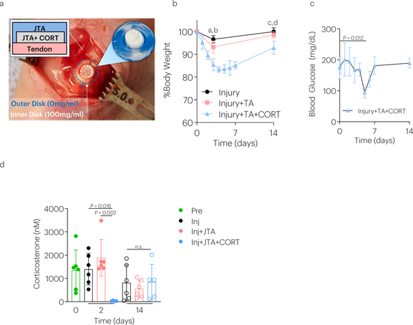
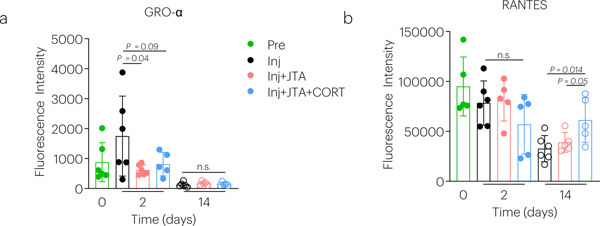
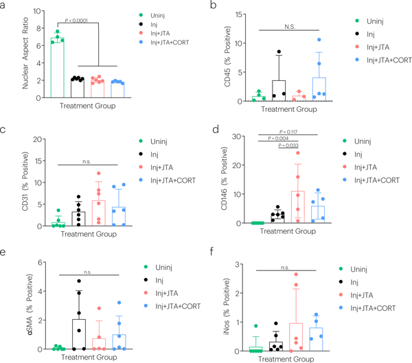
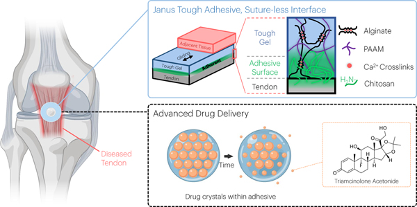
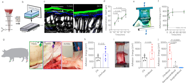
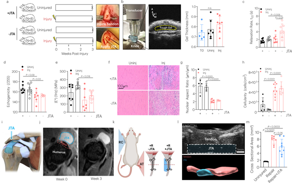
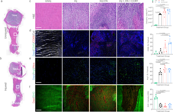
Hydrogels that provide mechanical support and sustainably release therapeutics have been used to treat tendon injuries. However, most hydrogels are insufficiently tough, release drugs in bursts, and require cell infiltration or suturing to integrate with surrounding tissue. Here we report that a hydrogel serving as a high-capacity drug depot and combining a dissipative tough matrix on one side and a chitosan adhesive surface on the other side supports tendon gliding and strong adhesion (larger than 1,000 J m-2) to tendon on opposite surfaces of the hydrogel, as we show with porcine and human tendon preparations during cyclic-friction loadings. The hydrogel is biocompatible, strongly adheres to patellar, supraspinatus and Achilles tendons of live rats, boosted healing and reduced scar formation in a rat model of Achilles-tendon rupture, and sustainably released the corticosteroid triamcinolone acetonide in a rat model of patellar tendon injury, reducing inflammation, modulating chemokine secretion, recruiting tendon stem and progenitor cells, and promoting macrophage polarization to the M2 phenotype. Hydrogels with 'Janus' surfaces and sustained-drug-release functionality could be designed for a range of biomedical applications.


Figures

ED Fig. 1
ED Fig. 1. Effect of CORT releasing JTAs on animal physiology over time.
(a) The JTA dissolution-controlled release system was surrounded by an outer JTA to stabilize it on the rat patellar tendon and enable a depot-based delivery system. (b) Rat body weight was examined over time. Data shown as mean ± s.d., as analyzed by ANOVAs, with post hoc tests with Bonferroni corrections. a: P = 0.0008, b: P = 0.002; c: P = 0.006; d: P = 0.0011. N=4–6 rats/group. (c) Blood glucose levels were evaluated over time. Data shown as mean ± s.d., as evaluated by a one-way ANOVA, with post hoc Tukey Tests for multiple comparisons. N=4–6 rats/group. (d) The effect of injury, JTA, and CORT on corticosterone levels 2 days and 14 days post-implantation. Data shown as mean ± s.d., as analyzed by a two-way repeated ANOVA (time and treatment), with post hoc Tukey Tests for multiple comparisons. N=4–6 samples/group.
ED Fig. 2
ED Fig. 2. Effect of the JTA and corticosteroid delivery on chemokines.
The effect of injury, JTA, and CORT on (a) GROα and (b) RANTES was evaluated after 2 and 14 days of healing. Data shown as mean ± s.d., as evaluated by a two-way repeated measures ANOVA with post hoc Tukey Tests for multiple comparisons. N=5–6 samples/group.
ED Fig. 3
ED Fig. 3. Effect of the JTA and corticosteroid delivery on tendon histological properties.
The effect of injury, JTA, and CORT on (a) nuclear aspect ratio, (b) CD45, (c) CD31, (d) CD146, (e) αSMA, and (f) iNos staining was evaluated after 2-weeks of healing. Data shown as mean ± s.d., as evaluated by a one-way ANOVA with post hoc Tukey Tests for multiple comparisons. N=4–6 tendons/group.
Fig. 1 |
Fig. 1 |. An overview of the vision for the multifunctional performance of the Janus Tough Adhesives (JTAs) for tendon.
JTA hydrogels were engineered that comprise a tough hydrogel dissipative matrix and adhesive chitosan surface. The tough hydrogel was synthesized by combining alginate ionically crosslinked with calcium and covalently crosslinked polyacrylamide (PAAM). Beyond tissue adhesion, the material can promote gliding of surrounding tissues and serve as a drug delivery system for local release of agents such as triamcinolone acetonide as exemplified in the present study.
Fig. 2 |
Fig. 2 |. The JTA adheres strongly to diverse tendon surfaces.
(a) Thin tendon planks for adhesion testing were made from bovine tendon samples using a cryotome. (b) The penetration of the chitosan bridging polymer into tendon in vitro was analyzed via placement of the JTA, using fluorescently labeled chitosan, on tendon planks, and quantifying chitosan depth over time. (c) Representative images and (d) quantification of penetration depth of chitosan into tendon over time. Mean values are shown and error bars represent ± s.d. (n=3 samples/group). Data were analyzed by a one-way ANOVA with post hoc t-tests with Bonferroni correction. (e) Peel testing was used to evaluate the adhesion energy (Blue: Tough Gel; Green: Adhesive Surface; White: tendon). (f) Quantification of the adhesion energy of JTA to tendon, over time, and comparison to the value achieved with TISSEEL at 100 minutes (dotted line). Mean values are shown and error bars represent ± s.d. (n=3 samples/group), (g) Adhesion of the JTA to wet and bloody porcine patellar, flexor carpi ulnaris, and Achilles tendons. (h) Porcine Achilles tendon immediately post-mortem was evaluated (JTA+Fresh) and compared to frozen tendon planks (JTA). Mean values are shown and error bars represent ± s.d. (n=6–7/group), as analyzed by a Student’s t-test. (i) The effect of blood on JTA adhesion to tendon was evaluated. Mean values are shown and error bars represent ± s.d. (n=6–7/group), as analyzed by a Student’s t-test. (j) Adhesion comparison of the JTA, Dermabond, and Surgicel to bloody tendons was evaluated. Mean values are shown and error bars represent ± s.d. (n=4–7 samples/group), as analyzed by a one-way ANOVA with post hoc t-tests with Bonferroni correction.
Fig. 3 |
Fig. 3 |. The JTA promotes tendon gliding.
(a,b) To evaluate friction properties during cyclic loading, an 8N normal force was applied vertically while a lower horizontal actuator moved cyclically +/− 10 mm (2 mm/s). (c) Images of the cyclic friction experiments for the JTA-JTA and tendon-tendon tests. (d) The effect of time and group on the coefficient of kinetic friction (μk) was evaluated. Mean values are shown and error bars represent ± s.d. (n=3–5 samples/group), as analyzed by a two-way ANOVA with post hoc tests with Bonferroni Corrections. (e,f) The ability for the JTA to support gliding of the flexor digitorum profundus tendon through the transverse carpal ligaments and over volar plates was evaluated in human cadaveric limbs.
Fig. 4 |
Fig. 4 |. The JTA is biocompatible to tendon and supports healing.
(a) The effect of patellar tendon injury and JTA implantation on tendon structural and mechanical properties was investigated. Following injury, the JTA was applied on the central midsubstance of the patellar tendon. (b) At 3-weeks post implantation, gel thickness was evaluated using high frequency ultrasound. Mean values are shown and error bars are ± s.d. (n=5–8 samples/group), as analyzed by a one-way ANOVA. A- Anterior; I- Inferior; M- Medial. (c-e) The effect of healing and JTA implantation on patellar tendon relaxation half-life, echogenicity, and |E*| was evaluated. Mean values are shown and error bars are +s.d. (n=6–8 samples/group), as analyzed by a two-way ANOVA with post hoc t-tests with Bonferroni corrections. (f-h) The effect of healing and JTA implantation on patellar tendon nuclear aspect ratio and cellularity was evaluated. Scale bar = 100 μm. Mean values are shown and error bars are ± s.d. (n=4–8 samples/group), as analyzed by a two-way ANOVA with post hoc t-tests with Bonferroni correction. (i,k) The JTA was designed to be placed over the supraspinatus tendon and pass under the acromion for testing in the rotator cuff. (j) T2-weighted MRI images were acquired from the rotator cuff to examine JTA placement and material compatibility over time. (k) The effect of the JTA on Achilles tendon healing in a repair model was evaluated. (l) Sagittal high frequency ultrasound imaging evaluated JTA (blue) placement over time adjacent to the Achilles tendon (red). (m) Axial high frequency ultrasound imaging examined the effect of tendon repair with and without the JTA on Achilles tendon cross sectional area. Mean values are shown and error bars are ± s.d. (n=8–9 tendons/group), as analyzed by a one-way ANOVA with post hoc t-tests with Bonferroni corrections.
Fig. 5 |
Fig. 5 |. The JTA enables dissolution-controlled release at high drug loadings.
(a) The CORT diffusion through the JTA was evaluated using a Franz cell. (b) Computational modeling and experiments evaluated the diffusion constant of the JTA. Mean values are shown and error bars are ± s.d. (n=4–6 samples/group). (c) In addition to diffusion-controlled release, dissolution-controlled release was investigated by loading CORT crystals (orange) within the JTA. (d) CORT aggregation and dissolution within the JTA was modeled based on brightfield microscopy images within pre-gel solution. (e,f) Dissolution of solid CORT was modeled using FE simulations under well mixed conditions to predict dissolution of particles. (g) Dissolution of CORT particles occurred from the gel periphery to center. (h) The effect of CORT loading on drug release was evaluated. Mean values are shown and error bars are ± s.d. (n=3 samples/group). (i,j) The effects of drug loading on JTA tensile properties and adhesion were evaluated. Mean values are shown and error bars are +s.d. (n=4 samples/group), as analyzed by a one-way ANOVA.
Fig. 6 |
Fig. 6 |. The JTA modulates vascularization and tendon properties in a rat model of patellar tendon injury.
(a) The effect of the JTA loaded with triamcinolone acetonide (CORT) on patellar tendon vascularity was investigated. (b) CORT concentrations in serum were evaluated over time in uninjured and injured groups. (c) High frequency ultrasound (HFUS) imaging revealed continued adhesion of the JTAs to tendon. (d,e) The effect of treatment and time on total blood vessel volume after injury was evaluated using HFUS Doppler imaging. Mean values are shown and error bars are ± s.d. (n=4–7 samples/group), as analyzed by a two-way ANOVA with post hoc t-tests with Bonferroni correction. (f,g,h) The effect of treatment on tendon biomechanics after injury was evaluated. Mean values are shown and error bars are ± s.d (n=6–9 samples/group), as analyzed by a one-way ANOVA with post hoc t-tests with Bonferroni correction.
Fig. 7 |
Fig. 7 |. The JTA induces immune modulation in tendon following injury.
(a,b) Full length histology (H&E staining) of uninjured and injured coronal tendon sections showing the region of interest (ROI) analyzed during image processing (midstance injury site) (c-f) Effect of injury, JTA, and JTA+CORT on tendon, as assessed by H&E (cellularity), immunostaining (CD68, Arg1), and SHG imaging (collagen signal) of tendon sections. Mean values are shown and error bars represent ± s.d. (n=5–6 samples/group), as analyzed by a one-way ANOVAs with post hoc Tukey Tests.

Similar articles

Cited by

References

    1. Surgeons A. A. o. O. Optimizing the Management of Rotator Cuff Problems. (2013). - PubMed
    1. Iannotti JP Full-Thickness Rotator Cuff Tears: Factors Affecting Surgical Outcome. J Am Acad Orthop Surg 2, 87–95 (1994). - PubMed
    1. Goutallier D, Postel JM, Gleyze P, Leguilloux P & Van Driessche S Influence of cuff muscle fatty degeneration on anatomic and functional outcomes after simple suture of full-thickness tears. J Shoulder Elbow Surg 12, 550–554, doi:10.1016/S1058274603002118 (2003). - DOI - PubMed
    1. McCarron JA et al. Failure with continuity in rotator cuff repair “healing”. Am J Sports Med 41, 134–141, doi:10.1177/0363546512459477 (2013). - DOI - PubMed
    1. Rodeo SA Biologic augmentation of rotator cuff tendon repair. J Shoulder Elbow Surg 16, S191–197, doi:10.1016/j.jse.2007.03.012 (2007). - DOI - PubMed

Publication types