[Preprint]. 2021 Dec 29:rs.3.rs-1211792.
doi: 10.21203/rs.3.rs-1211792/v1.

The SARS-CoV-2 B.1.1.529 Omicron virus causes attenuated infection and disease in mice and hamsters

Affiliations

The SARS-CoV-2 B.1.1.529 Omicron virus causes attenuated infection and disease in mice and hamsters

Michael Diamond et al. Res Sq. .

Update in

  • SARS-CoV-2 Omicron virus causes attenuated disease in mice and hamsters.
    Halfmann PJ, Iida S, Iwatsuki-Horimoto K, Maemura T, Kiso M, Scheaffer SM, Darling TL, Joshi A, Loeber S, Singh G, Foster SL, Ying B, Case JB, Chong Z, Whitener B, Moliva J, Floyd K, Ujie M, Nakajima N, Ito M, Wright R, Uraki R, Warang P, Gagne M, Li R, Sakai-Tagawa Y, Liu Y, Larson D, Osorio JE, Hernandez-Ortiz JP, Henry AR, Ciuoderis K, Florek KR, Patel M, Odle A, Wong LR, Bateman AC, Wang Z, Edara VV, Chong Z, Franks J, Jeevan T, Fabrizio T, DeBeauchamp J, Kercher L, Seiler P, Gonzalez-Reiche AS, Sordillo EM, Chang LA, van Bakel H, Simon V; Consortium Mount Sinai Pathogen Surveillance (PSP) study group; Douek DC, Sullivan NJ, Thackray LB, Ueki H, Yamayoshi S, Imai M, Perlman S, Webby RJ, Seder RA, Suthar MS, García-Sastre A, Schotsaert M, Suzuki T, Boon ACM, Diamond MS, Kawaoka Y. Halfmann PJ, et al. Nature. 2022 Mar;603(7902):687-692. doi: 10.1038/s41586-022-04441-6. Epub 2022 Jan 21. Nature. 2022. PMID: 35062015 Free PMC article.

Abstract

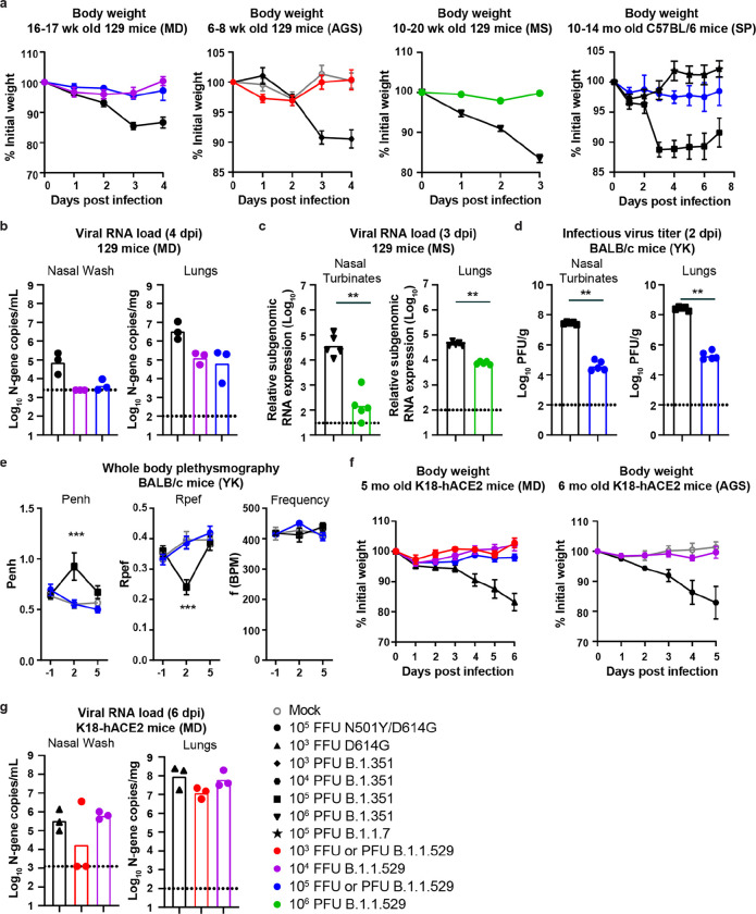
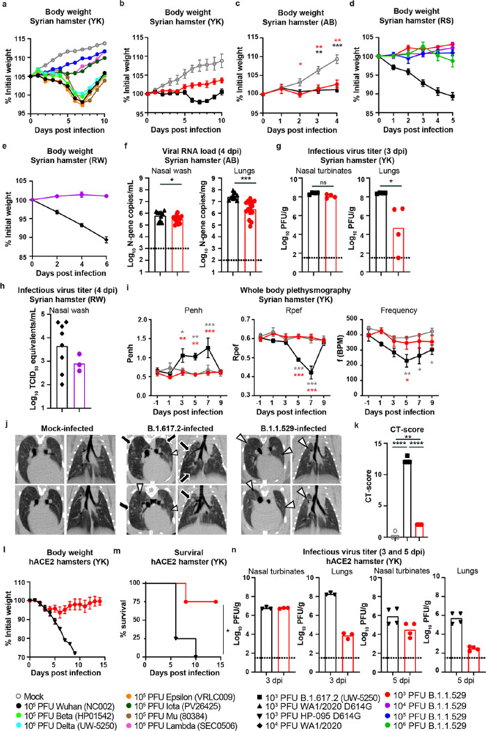
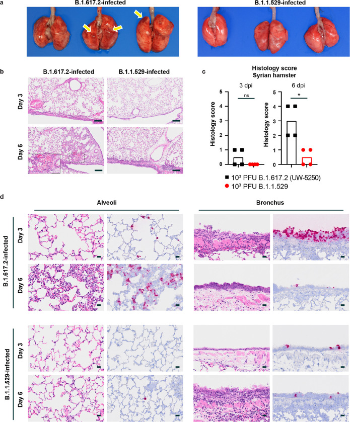
Despite the development and deployment of antibody and vaccine countermeasures, rapidly-spreading SARS-CoV-2 variants with mutations at key antigenic sites in the spike protein jeopardize their efficacy. The recent emergence of B.1.1.529, the Omicron variant1,2, which has more than 30 mutations in the spike protein, has raised concerns for escape from protection by vaccines and therapeutic antibodies. A key test for potential countermeasures against B.1.1.529 is their activity in pre-clinical rodent models of respiratory tract disease. Here, using the collaborative network of the SARS-CoV-2 Assessment of Viral Evolution (SAVE) program of the National Institute of Allergy and Infectious Diseases (NIAID), we evaluated the ability of multiple B.1.1.529 Omicron isolates to cause infection and disease in immunocompetent and human ACE2 (hACE2) expressing mice and hamsters. Despite modeling and binding data suggesting that B.1.1.529 spike can bind more avidly to murine ACE2, we observed attenuation of infection in 129, C57BL/6, and BALB/c mice as compared with previous SARS-CoV-2 variants, with limited weight loss and lower viral burden in the upper and lower respiratory tracts. Although K18-hACE2 transgenic mice sustained infection in the lungs, these animals did not lose weight. In wild-type and hACE2 transgenic hamsters, lung infection, clinical disease, and pathology with B.1.1.529 also were milder compared to historical isolates or other SARS-CoV-2 variants of concern. Overall, experiments from multiple independent laboratories of the SAVE/NIAID network with several different B.1.1.529 isolates demonstrate attenuated lung disease in rodents, which parallels preliminary human clinical data.


Figures

Figure 1
Figure 1. B.1.1.529 is attenuated in different mouse models of SARS-CoV-2 infection.
a, Far left: Weight change in 129 mice inoculated via intranasal route with 104 (purple circles, n = 6) or 105 (blue circles, n = 6) FFU of B.1.1.529 (strain hCoV-19/USA/WI-WSLH-221686/2021) or 105 FFU of WA1/2020 N501Y/D614G (black circles, n = 6). Center left: Weight change in mock-infected 129 mice (grey circles, n = 4) or 129 mice inoculated intranasally with 103 (red circles, n = 5) PFU of B.1.1.529 (strain hCoV-19/USA/NY-MSHSPSP-PV44476/2021) or B.1.351 variant of SARS-CoV-2 (black diamonds, n = 3). Center right: Weight change in 129 mice inoculated intranasally with 106 PFU of B.1.1.529 (strain hCoV19/EHC_C19_2811C, green circles, n = 5) or B.1.351 (black triangles, n = 5). Far right: Weight change in 10 to 14-month-old C57BL/6 mice inoculated intranasally with 105 PFU of B.1.1.529 (strain hCoV19/EHC_C19_2811C, blue circles, n = 4), B.1.1.7 (black star, n = 10), or B.1.351 (black squares, n = 18). Data are mean ± SEM. b, Nasal wash and lung viral RNA levels in 129 mice inoculated with 104 (purple circles, n = 3) or 105 (blue circles, n = 3) FFU of B.1.1.529 (strain hCoV-19/USA/WI-WSLH-221686/2021) or 105 FFU of WA1/2020 N501Y/D614G (black circles, n = 3). c, Nasal turbinates and lung viral RNA levels in 129 mice inoculated with 106 PFU of B.1.1.529 (strain hCoV19/EHC_C19_2811C, green circles, n = 5) or B.1.351 (black triangles, n = 5) (** P < 0.01, by Mann-Whitney U test). d, Nasal turbinates and lung virus titers from BALB/c mice inoculated with 105 PFU of B.1.1.529 (strain hCoV-19/Japan/NC928–2N/2021, blue circles, n = 5) or B.1.351 (black squares, n = 5) (** P < 0.001, by Mann-Whitney U test). e, Pulmonary function analysis in infected BALB/c mice. Penh, a surrogate marker for bronchoconstriction or airway obstruction, was measured by whole body plethysmography. Data are presented as the mean ± SEM. P values were calculated by using pairwise comparisons after a linear mixed model analysis (***P < 0.001). Asterisks indicate statistically significant differences between B.1.351-infected (n = 5) and B.1.1.529-infected (n = 5) or uninfected animals (n = 5). f, Left: Weight change in 5-month-old K18-hACE2 transgenic mice inoculated intranasally with 103 (red circles, n = 3), 104 (purple circles, n = 6), or 105 (blue circles, n = 3) FFU of B.1.1.529 (strain hCoV-19/USA/WI-WSLH-221686/2021) or 103 FFU of WA1/2020 D614G (black triangles, n = 6). Right: Weight change in 6-month-old K18-hACE2 transgenic mice inoculated intranasally with 104 (purple circles, n = 6) PFU of B.1.1.529 (strain hCoV-19/USA/NY-MSHSPSP-PV44476/2021), or 104 PFU of B.1.351 (black hexagon, n = 6)). Age-matched uninfected mice (grey circles, n = 4) were included as controls. Data are presented as the mean ± SEM. g, Nasal wash and lung viral RNA levels in K18-hACE2 mice inoculated with 103 FFU of WA1/2020 D614G (black triangles, n = 3), or 103 (red circles, n =3) or 104 (purple circles, n =3) FFU of B.1.1.529 (strain hCoV-19/USA/WI-WSLH-221686/2021). The results are from one experiment and each symbol in b, c, d, and g represents an individual animal. The dotted line is the limit of detection. Infection studies in panels a, b and f with WA1/2020 N501Y/D614G are shown as comparisons and were adapted from published data.
Figure 2
Figure 2. B.1.1.529 is attenuated in wild-type and human ACE2-transgenic Syrian hamsters.
a, Weight change in uninfected age-matched Syrian hamsters (open grey circles, n = 4) or in hamsters inoculated intranasally with 105 PFU (n = 9) of B.1.1.529 (strain hCoV-19/Japan/NC928–2N/2021, blue circles), B.1.351 (Beta, HP01542), B.1.617.2 (Delta, strain hCoV-19/USA/WI-UW-5250/2021), B.1.621 (Mu, 80384), B.1.427 (Epsilon, VRLC009), B.1.526 (Iota, PV26425), C.37 (Lambda, SEC0506), or Wuhan-1 (black circles). b, Weight change in uninfected age-matched Syrian hamsters (open grey circles, n = 3) or in hamsters inoculated intranasally with 103 PFU of B.1.1.529 (strain hCoV-19/Japan/NC928–2N/2021, red circles, n = 4) or B.1.617.2 (black squares, n = 4). Data are mean ± SEM. c, Weight change in uninfected age-matched Syrian hamsters (open grey circles, n = 9) or in hamsters inoculated intranasally with 103 PFU of B.1.1.529 (strain hCoV-19/USA/WI-WSLH-221686/2021, red circles, n = 10) or WA1/2020 D614G (black triangles, n = 6). Data are mean ± SEM. (* P < 0.05, ** P < 0.01, *** P < 0.001 by two-way ANOVA with a Dunnett’s correction). d, Weight change in Syrian hamsters inoculated intranasally with 103 (red circles, n = 4), 104 (purple circles, n = 4), 105 (blue circles, n = 4), or 106 (green circles, n = 4) PFU of B.1.1.529 (strain hCoV19/EHC_C19_2811C) or 103 PFU of B.1.617.2 (black squares, n = 4). Data are mean ± SEM. e, Weight change in hamsters inoculated intranasally with 104 PFU of B.1.1.529 (strain hCoV19/EHC_C19_2811C, purple circles, n = 5) or WA1/2020 (black diamonds, n = 9). Data are mean ± SEM. f, Nasal wash and lung viral RNA load in wild-type Syrian hamsters inoculated with 103 PFU of WA1/2020 D614G (black triangles, n = 15) or B.1.1.529 (strain hCoV-19/USA/WI-WSLH-221686/2021, red circles, n = 15) (*** P < 0.001, * P < 0.05 by unpaired t-test). g, Nasal turbinates and lung virus titer in wild-type Syrian hamsters inoculated with 103 PFU of B.1.617.2 (strain hCoV-19/USA/WI-UW-5250/2021, black squares, n = 4) or B.1.1.529 (strain hCoV-19/Japan/NC928–2N/2021, red circles, n = 4) (* P < 0.05, ns = not significant; Mann Whitney U test). h, Nasal wash viral RNA load (TCID50 equivalents/mL) in wild-type Syrian hamsters inoculated with 104 PFU of WA1/2020 (black diamonds, n = 8) or B.1.1.529 (strain hCoV19/EHC_C19_2811C, purple circles, n = 3). i, Pulmonary function analysis in infected Syrian hamsters. Penh was measured by whole body plethysmography. Data are mean ± SEM. P values were calculated by using pairwise comparisons after a linear mixed model analysis (*** P < 0.001, ** P < 0.01, * P < 0.05). Asterisks indicate statistically significant differences between B.1.617.2-infected (n = 4) and B.1.1.529-infected (n = 4) or uninfected animals (n = 3). j, Representative micro-CT axial and coronal images of the lungs of mock-infected (n = 3) or B.1.617.2- (n = 4) and B.1.1.529-infected (n = 4) hamsters on 7 dpi. Lung abnormalities included multifocal nodules (black arrows), ground glass opacity (white arrowheads), and regions of lung consolidation (white arrows) that were peripheral, bilateral, and multilobar. Pneumomediastinum is indicated with white asterisks. k, CT score for uninfected hamsters (open grey circles, n = 3) or those inoculated with 103 PFU of B.1.617.2 (black squares, n = 4) or B.1.1.529 (strain hCoV-19/Japan/NC928–2N/2021, red circles, n = 4). l, Weight change in hACE2-transgenic Syrian hamsters inoculated intranasally with 103 PFU of HP-095 D614G (black triangles, n = 4) or B.1.1.529 (strain hCoV-19/USA/WI-WSLH-221686/2021, red circles, n = 4). m, Survival analysis of hACE2-transgenic Syrian hamsters after inoculation with 103 PFU of HP-095 D614G (black triangles, n = 4) or B.1.1.529 (strain hCoV-19/USA/WI-WSLH-221686/2021, red circles, n = 4) (* P < 0.05; log-rank test). n, Nasal turbinate and lung infectious virus titer by plaque assay at 3 and 5 dpi from hACE2-transgenic Syrian hamsters inoculated with 103 PFU of HP-095 D614G (black triangles) or B.1.1.529 (strain hCoV19/USA/WI-WSLH-221686/2021, red circles); n = 3 (3 dpi), n = 4 (5 dpi). The results in the Figure are from one (a, b, d-e, and g-n) or two to three independent (c, and f) experiments. Each symbol represents an individual animal. Dotted lines represent the limit of detection.
Figure 3
Figure 3. Pathological findings in the lungs of SARS-CoV-2 infected Syrian hamsters.
The hamsters were infected with B.1.617.2 or B.1.1.529 variant of SARS-CoV-2 and sacrificed on 3 days (n = 4) and 6 days (n = 4) post infection for histopathological examinations. a, Macroscopic images of the lungs obtained from B.1.617.2 or B.1.1.529-infected Syrian hamsters at 6 dpi. Yellow arrows indicate areas of pulmonary hemorrhage. b, Representative histopathological images for the lung sections obtained from the animals infected with B.1.617.2 or B.1.1.529 at 3 or 6 dpi are shown at low magnification. Scale bars, 200 μm. Focal alveolar hemorrhage found in B.1.617.2-infected animals at 6 dpi is highlighted by dashed lines and shown at higher magnification in the inset image (scale bar indicating 100 μm). c, Histopathological score of viral pneumonia in the lungs of infected hamsters. Score was determined based on the percentage of alveolitis in a given section collected from each animal in each group using the following scoring: 0, no pathological change; 1, affected area (≤10%); 2, affected area (<50%, >10%); 3, affected area (≥50%); an additional point was added when pulmonary edema and/or alveolar hemorrhage was observed. Data are expressed as scatter plots with the median score ± 95% confidential interval. Each dot represents the score of each animal. Comparison between B.1.617.2 (n = 4) or B.1.1.529 (n = 4) was performed using Kruskal-Wallis test with a Dunn’s post-tests. ns, not significant; *, P < 0.05. d, RNA in situ hybridization (RNA-ISH) for SARS-CoV-2 viral RNA. Representative RNA-ISH images for the alveoli and bronchi of hamsters infected with B.1.617.2 (n = 4) or B.1.1.529 (n = 4) virus at 3 or 6 dpi are shown. Left panels, alveolar region. Right panels, bronchial region. Scale bars, 20 μm.

References

    1. Callaway E. & Ledford H. How bad is Omicron? What scientists know so far. Nature, doi:10.1038/d41586-021-03614-z (2021). - DOI - PubMed
    1. Torjesen I. Covid-19: Omicron may be more transmissible than other variants and partly resistant to existing vaccines, scientists fear. BMJ (Clinical research ed 375, n2943, doi:10.1136/bmj.n2943 (2021). - DOI - PubMed
    1. Muñoz-Fontela C. et al. Animal models for COVID-19. Nature 586, 509–515, doi:10.1038/s41586-020-2787-6 (2020). - DOI - PMC - PubMed
    1. Letko M., Marzi A. & Munster V. Functional assessment of cell entry and receptor usage for SARS-CoV-2 and other lineage B betacoronaviruses. Nature microbiology 5, 562–569, doi:10.1038/s41564-020-0688-y (2020). - DOI - PMC - PubMed
    1. Pinto D. et al. Cross-neutralization of SARS-CoV-2 by a human monoclonal SARS-CoV antibody. Nature 583, 290–295, doi:10.1038/s41586-020-2349-y (2020). - DOI - PubMed

Publication types

LinkOut - more resources