Review
. 2022 Jan 1;11(1):e210024.
doi: 10.1530/ETJ-21-0024.

Cellular and molecular basis of thyroid autoimmunity

Affiliations
Review

Cellular and molecular basis of thyroid autoimmunity

Joanna Bogusławska et al. Eur Thyroid J. .

Abstract

Autoimmune thyroid disease (AITD) is the most common human autoimmune disease. The two major clinical manifestations of AITD are Graves' disease and Hashimoto's thyroiditis (HT). AITD is characterized by lymphocytic infiltration of the thyroid gland, leading either to follicular cell damage, thyroid gland destruction, and development of hypothyroidism (in HT) or thyroid hyperplasia, induced by thyroid antibodies which activate thyrotropin receptor (TSHR) on thyrocytes, leading to hyperthyroidism. The aim of this review is to present up-to-date picture of the molecular and cellular mechanisms that underlie the pathology of AITD. Based on studies involving patients, animal AITD models, and thyroid cell lines, we discuss the key events leading to the loss of immune tolerance to thyroid autoantigens as well as the signaling cascades leading to the destruction of thyroid gland. Special focus is given on the interplay between the environmental and genetic factors, as well as ncRNAs and microbiome contributing to AITD development. In particular, we describe mechanistic models by which SNPs in genes involved in immune regulation and thyroid function, such as CD40, TSHR, FLT3, and PTPN22, underlie AITD predisposition. The clinical significance of novel diagnostic and prognostic biomarkers based on ncRNAs and microbiome composition is also underscored. Finally, we discuss the possible significance of probiotic supplementation on thyroid function in AITD.

Keywords: AITD; Graves’ disease; Hashimoto’s thyroiditis; autoimmune thyroid disease; circRNAs; lncRNAs; miRNAs; microbiome; non-coding RNAs; thyroid antigens; thyroid autoimmunity.


Figures

Figure 1
Figure 1
Localization and physiological function of thyroid antigens in thyrocytes. The sodium iodide symporter (NIS) transports I and Na+ through the basolateral plasma membrane of a thyroid epithelial cell. The Na+/K+ ATPase pump maintains the sodium diffusion gradient required for NIS function. Pendrin participates in the apical iodide efflux into the colloid of thyroid follicle. Thyroid peroxidase (TPO) catalyzes iodination of tyrosines in thyroglobulin (Tg), which attaches one or two iodine to form monoiodotyrosine (MIT) or diiodotyrosine (DIT), respectively. TPO also catalyzes the coupling of iodotyrosine residues to form triiodothyronine (T3) and thyroxine (T4) attached to Tg, whereas the dual oxidase (DUOX) supplies hydrogen peroxide (H2O2) for thyroid hormones (TH) biosynthesis. Release of TH requires engulfing colloid material (endocytosis) to form intracellular endosomes (not shown) that merge with lysosomes to form an endolysosome. TH liberated from the Tg scaffold are subsequently secreted into the blood vessels. Binding of thyrotropin (TSH; or thyroid-stimulating antibody) to TSH receptor (TSHR) activates intracellular signaling by the cAMP pathway leading to thyrocytes growth, differentiation, as well as production and release of TH.
Figure 2
Figure 2
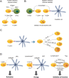
The mechanisms of selection, differentiation, and activation of T lymphocytes. (A) The positive selection of T cells (called thymocytes) expressing both CD4 and CD8 molecules in thymus cortex. Random recombination of genes encoding T cell receptor (TCR) peptides in T cell precursors leads to the expression of multiple TCR variants. TCR receptors interact with antigens presented by cortical thymic epithelial cells (cTECs) in complex with major histocompatibility complex (MHC) molecules. This interaction triggers the process of lymphocyte differentiation, leading to the generation of T cells expressing either CD4 or CD8 glycoprotein receptors on their surface. (B) The unwanted effect of TCR genes’ recombination is the production of receptor variants which recognize epitopes normally expressed by healthy cells (autoantigens). T cells which express autoreactive TCR variants are eliminated during the process of negative selection. The T CD4+ or T CD8+ cells resulting from positive selection migrate to the thymus medulla and interact with autopeptides complexed with MHC expressed at the surface of medullary thymic epithelial cells (mTECs) and dendritic cells. This interaction triggers the process of apoptotic deletion of autoreactive T cells. Negative selection is enabled by the ability of mTECs to express peptides (tissue-restricted antigens (TRAs) which are specific for different tissues of the organism (26, 27, 28). The expression of TRAs in mTEC is regulated by transcription factors, Aire and Fezf2. (C) Differentiation of T CD4+ cells. Following the process of central selection, T cells which do not react with autoantigens migrate to the lymphoid organs (spleen and lymph nodes) and then spread to all peripheral tissues. Depending on the signaling triggered by various cytokines, T CD4+ cells can differentiate to various types of T helper cells (Th) and T regulatory lymphocytes (Tregs) (28). (D) The mechanisms regulating activation and anergy of T lymphocytes. Activation of T lymphocytes: TCR expressed on T lymphocytes recognizes antigen complexed with MHC expressed by antigen-presenting cells (APC). T cell activation is triggered only in the presence of additional stimulation resulting from the interaction of CD28 and CD80/86 receptor. Anergy: In the absence of co-stimulatory signal, T cell undergoes anergy. The signals inhibiting T cell activity are generated by interaction between CTLA-4 and PD-1 receptors (on T cell surface) and their respective ligands (CD80/86 and PD-L1/2), expressed by APC. Inhibition: Tregs inhibit the functioning of T CD4+ and T CD8+ cells by triggering several mechanisms, including secretion of cytokines (interleukin-10 (IL-10), transforming growth factor B (TGFB), and IL-35) leading to inhibition of T cell proliferation, sequestration of IL-2, which triggers T cells’ apoptosis, as well as secretion of granzymes which exert cytotoxic effect on T cells. Tregs can also act directly by the molecules expressed on their cell surface which interact with T cell surface receptors leading to proliferation attenuation (30).
Figure 3
Figure 3
The mechanisms contributing to autoimmune thyroid disease (AITD) pathobiology. The mechanisms triggering the cascade of events leading to AITD involve the interplay between environmental factors (e.g. viral infection), epigenetic/genetic predispositions, and microbiome of which dysfunction contributes to the loss of immune tolerance, activation of autoreactive lymphocytes, and inflammation, leading to the damage of thyrocytes and clinical AITD.
Figure 4
Figure 4
The key mechanisms leading to the destruction of thyroid gland in Hashimoto’s thyroiditis (HT). APC activate T CD4+ lymphocytes which trigger their differentiation into T helper cells (Th1, Th2, and Th17). Th1 lymphocytes secreting mainly IL-12, TNFA, and INFG activate cytotoxic T lymphocytes (Tc) and macrophages which directly target and destroy thyroid follicular cells. The cytokines released by Th1 activate Tc, triggering apoptosis of thyrocytes induced by cytotoxins (perforin, granzymes, and granulysine) or FasL–Fas interaction. Thyroid glands of HT patients express high levels of Fas on the surface of follicular cells. Activated Tc express FasL, which interacts with Fas on thyrocytes, triggering pro-apoptotic signaling cascade. Th2 cells stimulate B cells leading to the formation of plasma cells which produce antibodies directed against thyroid autoantigens which bind thyroid autoantigens and induce thyrocyte apoptosis (mediated by antibody-dependent cytotoxicity or complement activation). Pro-inflammatory Th17 lymphocytes secrete IL-17 which stimulates macrophages, fibroblasts, and epithelial cells to produce cytokines, triggering apoptosis of thyrocytes. The suppressive actions of T regulatory lymphocytes are attenuated in AITD, preventing the counteraction of pro-inflammatory Th17 activity (7, 24).
Figure 5
Figure 5
The interference between environmental and genetic factors contributing to AITD development. (A) The GD-associated C allele of CD40 rs1883832 C/T SNP affects Kozak sequence resulting in increased translation efficiency of CD40 receptor. CD40 is expressed on immune cells (e.g. B lymphocytes) and thyrocytes. The environmental factors (e.g. viral infection or iodine excess) lead to the local inflammation, resulting in T cell-mediated CD40 activation, triggering cascades activating the expression of cytokines, including IL-6. The latter acts on B cells, leading to their activation and differentiation into plasma cells which secrete large amounts of thyroid-specific antibodies (40, 62). (B) The GD-associated rs12101261 TSHR SNP affects the binding site of promyelocytic leukemia zinc finger protein (PLZF) transcription factor which acts as a TSHR repressor. T allele of rs12101261 determines stronger binding of PLZF, thereby contributing to more efficient repression of TSHR transcription. IFNA (e.g. induced by viral infection) induces enrichment of histone H3 lysine 4-monomethylated (H3K4me1) at the site of rs12101261 and augments PLZF-mediated TSHR repression. This in turn leads to decreased intrathymic TSHR expression which facilitates loss of immune tolerance by T lymphocytes underexposed to the thyroid autoantigen (34). (C) FLT3 encodes fms-related tyrosine kinase 3 which acts as a receptor involved in functioning of dendritic cells and responses to viral infections. The HT-linked rs76428106-C allele of FLT3 creates a cryptic splice site which introduces the premature stop codon, potentially leading to the synthesis of the receptor devoid of the kinase domain. The expression of the rs76428106-C variant is associated with >20% decrease of the WT FLT3 and nearly two-fold increase of the FLT3 ligand. The resulting increase of the FLT3 ligand level leads to overactivation of the full-length receptor, contributing to the preferential expansion of plasmocytoid dendritic cells which in response to viral infection produce large amounts of interferon. The same study also confirmed that this variant was predisposing to other autoimmune diseases (i.e. systemic lupus erythematosus, rheumatoid-factor/anti-CCP-positive rheumatoid arthritis, and coeliac disease) (41).
Figure 6
Figure 6
The mechanisms by which PTPN22 polymorphism may contribute to AITD development. PTPN22 encodes lymphoid tyrosine phosphatase (Lyp) which dephosphorylates Src family of kinases. WT Lyp affects the functioning of B cells (left panel) and T cells (right panel) by inhibiting the signaling triggered by BCR and TCR, respectively. A 1858C/T SNP leads to R620W substitution, resulting in LypR620W variant which attenuates BCR and TCR signaling more strongly than the WT counterpart. It is proposed that dysfunction of Lyp can lead to inefficient deletion of autoreactive B cells. The WT Lyp is recruited to plasma membrane where it interacts with C-terminal Src kinase (Csk). Following TCR stimulation, Lyp dissociates from Csk and is recruited to lipid rafts to dephosphorylate its substrates. R620W substitution impairs Csk binding, leading enhanced LypR620W recruitment to lipid rafts and stronger inhibition of TCR signaling. In T cells, apart from inhibition of TCR cascade, LypR620W enhances the signaling triggered by CD28, resulting in Akt/mTOR activation. These alterations lead to the attenuation of the suppressive Treg actions which cannot prevent Th1 responsiveness, leading to the increased production of IFNG. On the other hand, the responsiveness of Th17 lymphocytes is reduced, as reflected by decreased IL-17 production. LypR620W mutant contributes to the enhanced adhesion, migration, and homing of T cells by augmenting the signaling induced by lymphocyte function-associated antigen–1 (LFA-1), an integrin receptor regulating the adhesion and migration of T cells. The efficient attenuation of LFA-1 signaling is mediated by Lyp–Csk complexes. Since LypR620W does not interact with Csk, it cannot inhibit LFA-1 signaling (42, 63, 64, 65).
Figure 7
Figure 7
The mechanisms of ncRNA-mediated regulation of gene expression. (1.) LncRNA can affect gene expression by influencing chromatin regulation or the functioning of transcription factors. (2.) LncRNA and (3.) miRNA affect mRNA translation and/or promote mRNA degradation. (4.) LncRNA can act as scaffolds recruiting and binding proteins. (5.) LncRNA can influence posttranslational protein modifications such as phosphorylation. (6.) LncRNA and (7.) circRNA act as sponges binding miRNAs and precluding their regulatory effects.
Figure 8
Figure 8
The effects of dysbiosis on immune regulation and autoimmunity. Healthy microbiota produce compounds that act as ligands of TLR and NLR receptors (nucleotide oligomerization domain (NOD)-like receptors) expressed on immune cells, including macrophages and dendritic cells which release cytokines and metabolites such as TGFB, retinoic acid, and interleukins. The cytokines act on target cells, including Th17 lymphocytes and Tregs, ensuring the balance between pro-inflammatory and anti-inflammatory mechanisms. Dysbiosis leads to disturbed regulation of this signaling cascade, shifting the balance toward pro-inflammatory Th17, contributing to inflammation and autoimmunity (51, 53).

References

    1. Mclachlan SM, Rapoport B. Breaking tolerance to thyroid antigens: changing concepts in thyroid autoimmunity. Endocrine Reviews 20143559–105. (10.1210/er.2013-1055) - DOI - PMC - PubMed
    1. Tomer Y, Huber A. The etiology of autoimmune thyroid disease: a story of genes and environment. Journal of Autoimmunity 200932231–239. (10.1016/j.jaut.2009.02.007) - DOI - PMC - PubMed
    1. Wood LC, Ingbar SH. Hypothyroidism as a late sequela in patient with Graves’ disease treated with antithyroid agents. Journal of Clinical Investigation 1979641429–1436. (10.1172/JCI109601) - DOI - PMC - PubMed
    1. Eckstein A, Philipp S, Goertz G, Banga JP, Berchner-Pfannschmidt U. Lessons from mouse models of Graves’ disease. Endocrine 202068265–270. (10.1007/s12020-020-02311-7) - DOI - PMC - PubMed
    1. Wiersinga WM.Advances in treatment of active, moderate-to-severe Graves’ ophthalmopathy. Lancet: Diabetes and Endocrinology 20175134–142. (10.1016/S2213-8587(1630046-8) - DOI - PubMed