. 2022 Jan 1;78(Pt 1):91-103.
doi: 10.1107/S2059798321011785. Epub 2022 Jan 1.

Catalytically active holo Homo sapiens adenosine deaminase I adopts a closed conformation

Affiliations

Catalytically active holo Homo sapiens adenosine deaminase I adopts a closed conformation

Minh Thu Ma et al. Acta Crystallogr D Struct Biol. .

Abstract

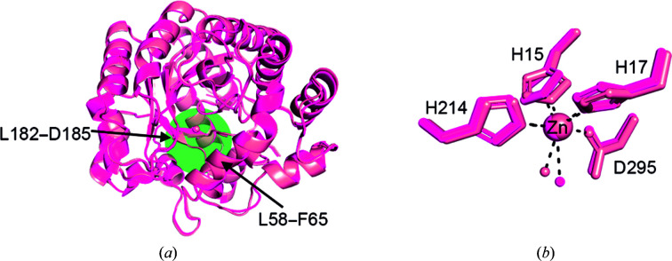
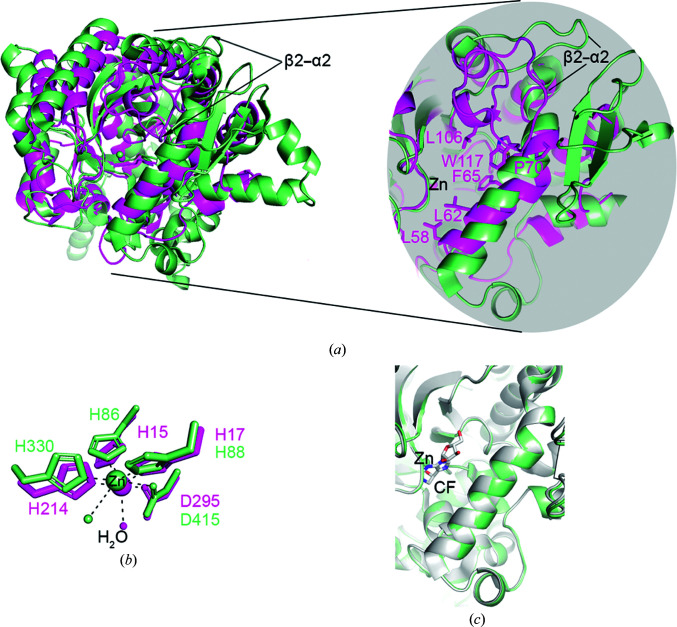
Homo sapiens adenosine deaminase 1 (HsADA1; UniProt P00813) is an immunologically relevant enzyme with roles in T-cell activation and modulation of adenosine metabolism and signaling. Patients with genetic deficiency in HsADA1 suffer from severe combined immunodeficiency, and HsADA1 is a therapeutic target in hairy cell leukemias. Historically, insights into the catalytic mechanism and the structural attributes of HsADA1 have been derived from studies of its homologs from Bos taurus (BtADA) and Mus musculus (MmADA). Here, the structure of holo HsADA1 is presented, as well as biochemical characterization that confirms its high activity and shows that it is active across a broad pH range. Structurally, holo HsADA1 adopts a closed conformation distinct from the open conformation of holo BtADA. Comparison of holo HsADA1 and MmADA reveals that MmADA also adopts a closed conformation. These findings challenge previous assumptions gleaned from BtADA regarding the conformation of HsADA1 that may be relevant to its immunological interactions, particularly its ability to bind adenosine receptors. From a broader perspective, the structural analysis of HsADA1 presents a cautionary tale for reliance on homologs to make structural inferences relevant to applications such as protein engineering or drug development.

Keywords: adenosine deaminase 1; closed conformation; homologs; structural inference.


Figures

Figure 1
Figure 1
Functional properties of purified HsADA1. (a) SDS–PAGE analysis of HsADA1 before (lane 1) and after (lane 2) cleavage of the His tag with TEV protease, showing the expected decrease in molecular mass. (b) Size-exclusion chromatography elution profile for cleaved HsADA1. (c) Michaelis–Menten kinetic parameters of cleaved HsADA1 at pH 7.4, which agree with previously reported values. (d) Stability of tagged and cleaved HsADA1 obtained from differential scanning fluorimetry. The melting temperature was determined as the inflection point of the first derivative with respect to temperature of the 330 nm:350 nm absorbance ratio. (e) His-tagged HsADA1 activity as a function of pH condition (n = 32 per pH condition). Due to the different concentrations of enzyme necessary to obtain a robust signal for each pH condition, the original rate calculation (determined by linear regression from the initial 10% of substrate degradation) in units of µM adenosine per second was divided by the concentration of HsADA1 in µM to obtain a normalized unit of s−1.
Figure 2
Figure 2
Structure of holo HsADA1. (a) Overlay of holo HsADA1 monomers in the crystal asymmetric unit. Monomer A (magenta) and monomer B (light pink) exhibit only minor structural differences, with an overall r.m.s.d. of 0.34 Å. The green circle indicates the structural gate formed by residues Leu58–Phe65 and Leu182–Asp185. (b) Enlarged view of the zinc-binding regions of monomers A and B. The side chains of the active-site residues are shown as sticks along with the coordinating water molecules. Dashed lines represent coordinating interactions with zinc.
Figure 3
Figure 3
Comparison of HsADA1 with other structures of mammalian ADA1 enzymes. (a) Overlay of holo HsADA1 monomer A (magenta) with the unpublished structure of substrate/Ni2+-bound HsADA1 (PDB entry 3iar, lime green). (b) Comparison of Zn2+ and Ni2+ coordination in HsADA1 and PDB entry 3iar, respectively. (c) Enlarged view of the C-terminal helix (residues 354–364) that is present in PDB entry 3iar but absent in HsADA1. (d) Overlay of holo HsADA1 monomer A (magenta) and holo BtADA (cyan) with enlarged views of the Zn2+ environment, C-terminal helix and structural gate. HsADA1 adopts an open conformation with shifts in Leu58, Leu62 and the Leu182–Asp185 loop compared with the closed conformation of holo BtADA. (e) Comparison of the structural gate of HsADA1 monomer A (magenta) with MmADA complexed with DCF (brown) and 1-deaza-adenosine (light blue) and with BtADA complexed with EHNA (orange) and FR235380 (lime green). HsADA1 has similar structural features at the catalytic gate to the closed conformation adopted by MmADA–inhibitor complexes, while displaying shifts in this region from the open conformation adopted by BtADA–inhibitor complexes. (f) Overlay of monomer A (magenta) with holo MmADA (yellow) with enlarged views of the Zn2+ environment, C-­terminal site and structural gate. The structural features of the catalytic gate of HsADA1 overlap closely with those of holo MmADA.
Figure 4
Figure 4
Crystal contact analysis. (a) Overlay of four key illustrative structures in the region of the structural gate helix comprising residues 58–65. In all four structures Tyr67 participates in crystal contacts and is in a similar position. (b) Crystal contacts for holo HsADA1 showing polar contacts with the Pro354 and Pro355 main-chain carbonyls. (c) Crystal contacts for holo MmADA showing a water-mediated contact with Glu345. (d) Crystal contacts for holo BtADA showing interaction between Tyr67 and Lys206 as well as between Asp61 and Ser207. (e) Crystal contacts in the closed-conformation structure of BtADA showing contact only between Tyr67 and the main-chain atoms of Val205 and Gly208. Dashed lines represent contacts between 2.6 and 3.5 Å.
Figure 5
Figure 5
Cavity and receptor-binding site analysis. (a) Left panel, overview of holo HsADA1 with solvent cavities shown in pale yellow. Right panel, enlargement of the structural gate. (b) Two orientations of the structural gate overlaid with PDB entry 1add in which 1-deaza-adenosine is bound to MmADA, showing good agreement with the cavity in HsADA1. (c) Surface representation of the expected CD26 and adenosine receptor binding sites of HsADA1.
Figure 6
Figure 6
Comparison of HsADA1 with HsADA2. (a) Superposition of monomer A (magenta) of HsADA1 and holo HsADA2 (light green; PDB entry 3lgd), with arrows indicating the location of the β2–α2 loop. Inset, enlarged view of the β2–α2 loop showing interaction of β2–α2 with the helix of the structural gate in HsADA1 but not in HsADA2. (b) Enlarged view of the Zn2+ coordination environment. (c) Enlarged view of the structural gate showing that nonligated HsADA2 and CF-bound HsADA2 (gray; PDB entry 3lgg) adopt the same conformation, which is analogous to the open conformation of BtADA (see Fig. 3 ▸ e).

References

    1. Bhaumik, D., Medin, J., Gathy, K. & Coleman, M. S. (1993). J. Biol. Chem. 268, 5464–5470. - PubMed
    1. Bolanos-Garcia, V. M. & Chayen, N. E. (2009). Prog. Biophys. Mol. Biol. 101, 3–12. - PubMed
    1. Bradford, K. L., Moretti, F. A., Carbonaro-Sarracino, D. A., Gaspar, H. B. & Kohn, D. B. (2017). J. Clin. Immunol. 37, 626–637. - PubMed
    1. Casañal, A., Lohkamp, B. & Emsley, P. (2020). Protein Sci. 29, 1069–1078. - PMC - PubMed
    1. Chan, B., Wara, D., Bastian, J., Hershfield, M. S., Bohnsack, J., Azen, C. G., Parkman, R., Weinberg, K. & Kohn, D. B. (2005). Clin. Immunol. 117, 133–143. - PubMed