. 2022 Apr;303(1):54-62.
doi: 10.1148/radiol.210407. Epub 2022 Jan 4.

Prediction of Upstaging in Ductal Carcinoma in Situ Based on Mammographic Radiomic Features

Affiliations

Prediction of Upstaging in Ductal Carcinoma in Situ Based on Mammographic Radiomic Features

Rui Hou et al. Radiology. 2022 Apr.

Abstract

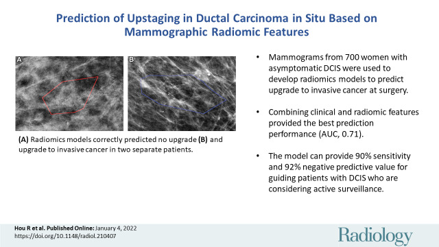
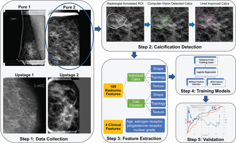
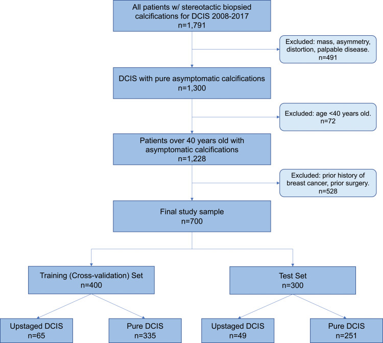
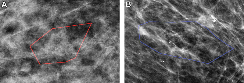
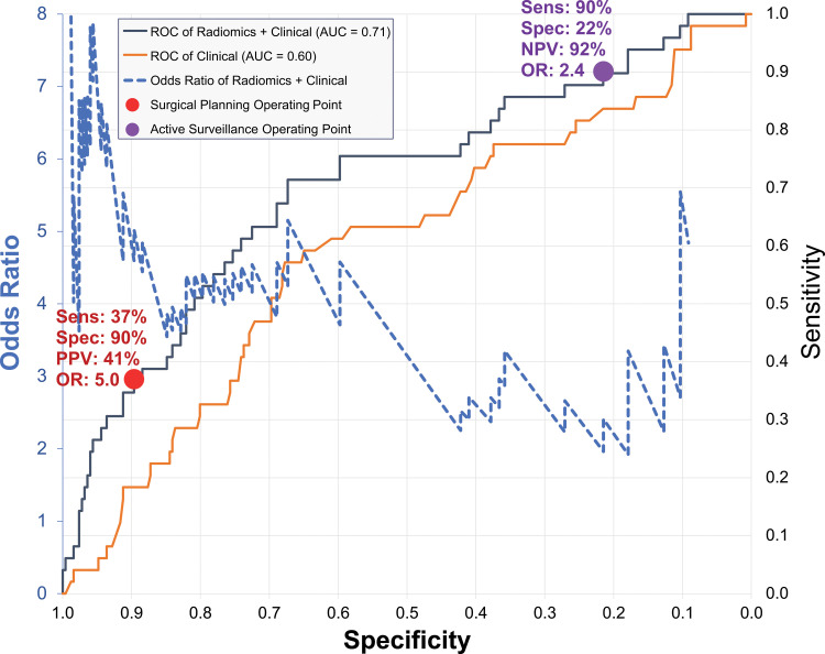
Background Improving diagnosis of ductal carcinoma in situ (DCIS) before surgery is important in choosing optimal patient management strategies. However, patients may harbor occult invasive disease not detected until definitive surgery. Purpose To assess the performance and clinical utility of mammographic radiomic features in the prediction of occult invasive cancer among women diagnosed with DCIS on the basis of core biopsy findings. Materials and Methods In this Health Insurance Portability and Accountability Act-compliant retrospective study, digital magnification mammographic images were collected from women who underwent breast core-needle biopsy for calcifications that was performed at a single institution between September 2008 and April 2017 and yielded a diagnosis of DCIS. The database query was directed at asymptomatic women with calcifications without a mass, architectural distortion, asymmetric density, or palpable disease. Logistic regression with regularization was used. Differences across training and internal test set by upstaging rate, age, lesion size, and estrogen and progesterone receptor status were assessed by using the Kruskal-Wallis or χ2 test. Results The study consisted of 700 women with DCIS (age range, 40-89 years; mean age, 59 years ± 10 [standard deviation]), including 114 with lesions (16.3%) upstaged to invasive cancer at subsequent surgery. The sample was split randomly into 400 women for the training set and 300 for the testing set (mean ages: training set, 59 years ± 10; test set, 59 years ± 10; P = .85). A total of 109 radiomic and four clinical features were extracted. The best model on the test set by using all radiomic and clinical features helped predict upstaging with an area under the receiver operating characteristic curve of 0.71 (95% CI: 0.62, 0.79). For a fixed high sensitivity (90%), the model yielded a specificity of 22%, a negative predictive value of 92%, and an odds ratio of 2.4 (95% CI: 1.8, 3.2). High specificity (90%) corresponded to a sensitivity of 37%, positive predictive value of 41%, and odds ratio of 5.0 (95% CI: 2.8, 9.0). Conclusion Machine learning models that use radiomic features applied to mammographic calcifications may help predict upstaging of ductal carcinoma in situ, which can refine clinical decision making and treatment planning. © RSNA, 2022.


Conflict of interest statement

Disclosures of conflicts of interest: R.H. No relevant relationships. L.J.G. Grants from ECOG/Acrin (TMIST) Alliance for Clinical Trials in Oncology Foundation AUR Breast Cancer Research Foundation AD Anderson Cancer Center; consulting fees from Hologic; payment for lectures from Medscape Reference; payment for expert testimony from Hare, Wynn, Newell and Newton LLP, leadership or fiduciary role in Society of Breast Imaging. M.A.M. No relevant relationships. J.R.M. Grant from University of Utah Subaward; participation on a Data Safety Monitoring board or advisory board at Duke University. L.M.K. No relevant relationships. C.C.M. Leadership or fiduciary role in International Society for Evolution, Ecology and Cancer. T.L. No relevant relationships. M.v.O. No relevant relationships. K.R. No relevant relationships. N.S. Participation on a Data Safety or advisory board at University of Exeter. M.W. No relevant relationships. J.T. No relevant relationships. J.W. Member Research Council KWF Dutch Cancer Society; Member Scientific Advisory Board Member Dutch Expert Center for Screening; Advisor for the population-based breast screening program by the National Institute for Public Health and the Environment on behalf of the Dutch Society of Pathology. E.S.H. Consulting fees from AstraZeneca; payment for lectures from Merck; participation on a Data Safety or advisory board at Duke University; leadership or fiduciary role from Immunis Clinetic; stock options from Clinetic. J.Y.L. Equipment grant from Nvidia.

Figures

None
Graphical abstract
Illustration of the study pipeline (step 1). A total of 700 women were
identified (step 2). Lesion annotations were masked by a breast radiologist,
and calcifications (calcs) were masked by a computer vision–based
algorithm and a deep learning–based U-net segmentation network (step
3). A total of 109 radiomic features and four clinical features were
collected (step 4). Models with those extracted features and training data
were trained (step 5). Selected models were validated on test data. NPV =
negative predictive value, OR = odds ratio, PPV = positive predictive value,
ROC = receiver operating characteristic, ROI = region of interest, sens =
sensitivity, spec = specificity.
Figure 1:
Illustration of the study pipeline (step 1). A total of 700 women were identified (step 2). Lesion annotations were masked by a breast radiologist, and calcifications (calcs) were masked by a computer vision–based algorithm and a deep learning–based U-net segmentation network (step 3). A total of 109 radiomic features and four clinical features were collected (step 4). Models with those extracted features and training data were trained (step 5). Selected models were validated on test data. NPV = negative predictive value, OR = odds ratio, PPV = positive predictive value, ROC = receiver operating characteristic, ROI = region of interest, sens = sensitivity, spec = specificity.
Study inclusion flowchart of patients with ductal carcinoma in situ
(DCIS).
Figure 2:
Study inclusion flowchart of patients with ductal carcinoma in situ (DCIS).
Mammographic images of patients with biopsy-proven ductal carcinoma in
situ (DCIS). (A) A 55-year-old woman (right magnification craniocaudal view)
diagnosed with DCIS only; model correctly classified as negative findings.
(B) A 64-year-old woman (left magnification mediolateral oblique view) with
DCIS at core biopsy but subsequently upstaged to invasive disease; model
correctly classified as positive findings. Red and blue polygons show
lesions annotated by the radiologist.
Figure 3:
Mammographic images of patients with biopsy-proven ductal carcinoma in situ (DCIS). (A) A 55-year-old woman (right magnification craniocaudal view) diagnosed with DCIS only; model correctly classified as negative findings. (B) A 64-year-old woman (left magnification mediolateral oblique view) with DCIS at core biopsy but subsequently upstaged to invasive disease; model correctly classified as positive findings. Red and blue polygons show lesions annotated by the radiologist.
Graph shows receiver operating characteristic (ROC) curves and odds
ratios (ORs) of prediction models. Receiver operating characteristic curves
are shown for two models: one using radiomic and clinical features (gray)
and one using clinical features alone (orange). Both receiver operating
characteristic curves are plotted as sensitivity (secondary vertical axis on
the right) versus specificity (horizontal axis). Blue dashed line is OR
curve, plotted as OR (primary vertical axis on left) versus specificity
(horizontal axis). Two operating points are shown with symbols and are
described in the text: high-sensitivity active surveillance (purple circle);
high-specificity surgical planning for sentinel node biopsy alongside with
lesion removal surgery (red circle). AUC = area under receiver operating
characteristic curve, NPV = negative predictive value, PPV = positive
predictive value, Sens = sensitivity, Spec = specificity.
Figure 4:
Graph shows receiver operating characteristic (ROC) curves and odds ratios (ORs) of prediction models. Receiver operating characteristic curves are shown for two models: one using radiomic and clinical features (gray) and one using clinical features alone (orange). Both receiver operating characteristic curves are plotted as sensitivity (secondary vertical axis on the right) versus specificity (horizontal axis). Blue dashed line is OR curve, plotted as OR (primary vertical axis on left) versus specificity (horizontal axis). Two operating points are shown with symbols and are described in the text: high-sensitivity active surveillance (purple circle); high-specificity surgical planning for sentinel node biopsy alongside with lesion removal surgery (red circle). AUC = area under receiver operating characteristic curve, NPV = negative predictive value, PPV = positive predictive value, Sens = sensitivity, Spec = specificity.

References

    1. Siegel RL , Miller KD , Jemal A . Cancer statistics, 2020 . CA Cancer J Clin 2020. ; 70 ( 1 ): 7 – 30 . - PubMed
    1. Ryser MD , Weaver DL , Zhao F , et al. . Cancer outcomes in DCIS patients without locoregional treatment . J Natl Cancer Inst 2019. ; 111 ( 9 ): 952 – 960 . - PMC - PubMed
    1. Sanders ME , Schuyler PA , Dupont WD , Page DL . The natural history of low-grade ductal carcinoma in situ of the breast in women treated by biopsy only revealed over 30 years of long-term follow-up . Cancer 2005. ; 103 ( 12 ): 2481 – 2484 . - PubMed
    1. Hieken TJ , Krishnamurthy V , Farolan M , Velasco JM . Long-term outcome of DCIS patients: p53 as a biomarker of ipsilateral recurrence . J Clin Oncol 2011. ; 29 ( 27_suppl ): 39 .
    1. Schulz S , Sinn P , Golatta M , et al. . Prediction of underestimated invasiveness in patients with ductal carcinoma in situ of the breast on percutaneous biopsy as rationale for recommending concurrent sentinel lymph node biopsy . Breast 2013. ; 22 ( 4 ): 537 – 542 . - PubMed

Publication types